Page 116 - TD-4-2
P. 116

Tumor Discovery                                                         Desmoplastic small round cell tumor


                                                               3 h after admission. No autopsy was conducted according
                                                               to the wishes of the patient’s family.
                                                               3. Discussion

                                                               DSRCTs were first reported by Gerald and Rosai in 1989.
                                                                                                             2
                                                               This condition occurs most frequently in young people,
                                                               and its peak incidence has been reported in people in
                                                               their 30s. It predominantly affects men and is exceedingly
                                                               rare in women. While it most frequently presents in the
                                                               peritoneum and omentum, occurrences at other sites,
                                                               including the pleura, ethmoid sinus, scalp, hands, posterior
                                                               cranial fossa, pancreas, ovaries, paratesticular region, and
                                                               kidneys, have been documented. 3-5
                                                                 Clinical symptoms of primary gynecological disease
                                                               are abdominal distension with abdominal pain and ascites,
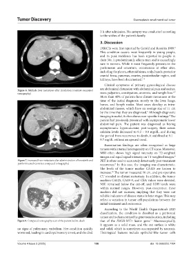
            Figure 6. Multiple liver metastases after irradiation (contrast computed
                                                                                                            6,7
            tomography)                                        mass palpation, constipation, anorexia, and weight loss.
                                                               More than 40% of patients have distant metastases at the
                                                               time of the initial diagnosis, mostly to the liver, lungs,
                                                               bones, and lymph nodes. Most cases develop as intra-
                                                               abdominal masses, which have an average size of 11 cm
                                                               by the time that they are diagnosed.  Although diagnostic
                                                                                            3
                                                               imaging is useful, it often shows non-specific findings.  The
                                                                                                         8
                                                               patient had previously presented with asymptomatic lower
                                                               abdominal pain. The patient was diagnosed as having
                                                               asymptomatic hypercalcemia; post-surgery, their serum
                                                               calcium levels decreased to 9.3 – 9.9 mg/dL, and during
                                                               the period from recurrence to death, it stabilized at 9.1 –
                                                               9.7 mg/dL without an upward trend.
                                                                 Examination findings are often recognized as large
                                                               tumors with internal heterogeneity on CT scans. Moreover,
                                                               MRI often shows high signal intensity on T2-weighted
                                                               images and equal signal intensity on T1-weighted images.
                                                                                                             8
            Figure 7. Increased liver metastasis after administration of lenvatinib and   PET is often used to accurately detect early post-treatment
            pembrolizumab (contrast computed tomography)       recurrence.  In this case, the imaging was characteristic.
                                                                        9
                                                               The levels of the tumor marker CA125 are known to
                                                               increase.  The tumor measured 10 cm, and pre-operative
                                                                      10
                                                               CT revealed no distant metastasis. In addition, the tumor
                                                               markers CA125, CA19-9, and CEA values were elevated,
                                                               NSE remained below the cut-off, and LDH levels were
                                                               within normal ranges. However, post-recurrence, these
                                                               markers did not increase, implying that they were not
                                                               reliable indicators of disease status in later stages. This may
                                                               reflect a variation in tumor cell populations between the
                                                               initial treatment and recurrence.
                                                                 According to the World Health Organization’s 2020
                                                               classification, the condition is classified as a peritoneal
                                                               tumor and is characterized by gene translocation, including
                                                                                              11
            Figure 8. Computed tomography scan of the patient before death  that of the  EWSR-WT1 fusion gene.  Macroscopically,
                                                               it appears as a solid mass, and the cut surface is white
            no signs of pulmonary embolism. Her condition quickly   and solid, which is sometimes accompanied by necrosis.
            worsened, leading to cardiopulmonary arrest, and she died   Histological features include epithelial-like tumor cells


            Volume 4 Issue 2 (2025)                        108                                doi: 10.36922/td.7104
   111   112   113   114   115   116   117   118   119   120   121