
Page 115 - TD-4-2
P. 115
Tumor Discovery Desmoplastic small round cell tumor
A C E
B D F
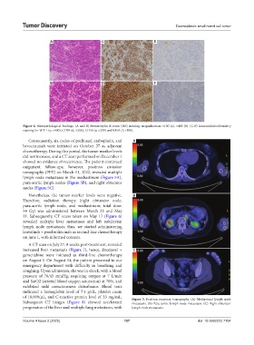
Figure 4. Histopathological findings. (A and B) Hematoxylin & eosin (HE) staining; magnification: ×100 (a), ×400 (b). (C-F) Immunohistochemistry
staining for WT-1 (c; ×400), CD99 (d; ×200), CD56 (e; ×200) and EMA (f; ×100).
Consequently, six cycles of paclitaxel, carboplatin, and A
bevacizumab were initiated on October 27 as adjuvant
chemotherapy. During this period, the tumor marker levels
did not increase, and a CT scan performed on December 1
showed no evidence of recurrence. The patient continued
outpatient follow-ups; however, positron emission
tomography (PET) on March 11, 2022, revealed multiple
lymph-node metastases in the mediastinum (Figure 5A),
para-aortic lymph nodes (Figure 5B), and right obturator
nodes (Figure 5C).
Nonetheless, the tumor marker levels were negative. B
Therefore, radiation therapy (right obturator node,
para-aortic lymph node, and mediastinum; total dose:
54 Gy) was administered between March 23 and May
11. Subsequently, CT scans taken on May 17 (Figure 6)
revealed multiple liver metastases and left subclavian
lymph node metastasis; thus, we started administering
lenvatinib + pembrolizumab as second-line chemotherapy
on June 1, with informed consent.
A CT scan on July 27, 8 weeks post-treatment, revealed
increased liver metastasis (Figure 7); hence, docetaxel + C
gemcitabine were initiated as third-line chemotherapy
on August 3. On August 14, the patient presented to our
emergency department with difficulty in breathing and
coughing. Upon admission, she was in shock, with a blood
pressure of 70/45 mmHg, requiring oxygen at 7 L/min
and SatO2 (arterial blood oxygen saturation) at 70%, and
exhibited mild consciousness disturbance. Blood tests
indicated a hemoglobin level of 7.1 g/dL, platelet count
of 18,000/μL, and C-reactive protein level of 33 mg/mL. Figure 5. Positron emission tomography. (A) Mediastinal lymph node
Subsequent CT images (Figure 8) showed accelerated metastasis. (B) Para-aortic lymph node metastasis. (C) Right obturator
progression of the liver and multiple lung metastases, with lymph node metastasis.
Volume 4 Issue 2 (2025) 107 doi: 10.36922/td.7104