
Page 114 - TD-4-2
P. 114
Tumor Discovery Desmoplastic small round cell tumor
2. Case presentation
Here, we report the case of a 33-year-old woman with three
pregnancies and three births. All procedures performed in
this study involving human participants were in accordance
with the ethical standards of the institutional and/or
national research committee and with the 1964 Helsinki
Declaration and its later amendments or comparable
ethical standards.
The patient gave birth in November 2020, and no
abnormalities were found during pregnancy or post-
partum examination. In October 2021, the patient
experienced frequent lower abdominal pain. She visited a
nearby obstetrics and gynecology clinic. Ultrasonography
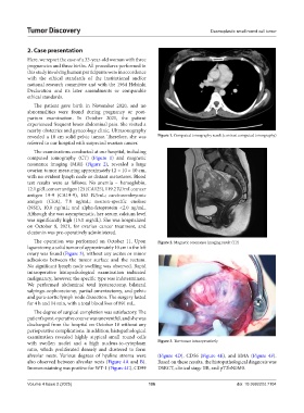
revealed a 10 cm solid pelvic tumor. Therefore, she was Figure 1. Computed tomography result (contrast computed tomography)
referred to our hospital with suspected ovarian cancer.
The examinations conducted at our hospital, including
computed tomography (CT) (Figure 1) and magnetic
resonance imaging (MRI) (Figure 2), revealed a large
ovarian tumor measuring approximately 12 × 10 × 10 cm,
with no evident lymph node or distant metastases. Blood
test results were as follows: No anemia – hemoglobin,
12.1 g/dL; cancer antigen 125 (CA125), 119.2 IU/mL; cancer
antigen 19-9 (CA19-9), 163 IU/mL; carcinoembryonic
antigen (CEA), 7.8 ng/mL; neuron-specific enolase
(NSE), 10.0 ng/mL; and alpha-fetoprotein <2.0 ng/mL.
Although she was asymptomatic, her serum calcium level
was significantly high (15.0 mg/dL). She was hospitalized
on October 8, 2021, for ovarian cancer treatment, and
elcatonin was pre-operatively administered.
The operation was performed on October 11. Upon Figure 2. Magnetic resonance imaging result (T2)
laparotomy, a solid tumor of approximately 10 cm in the left
ovary was found (Figure 3), without any ascites or minor
adhesions between the tumor surface and the rectum.
No significant lymph node swelling was observed. Rapid
intraoperative histopathological examination indicated
malignancy; however, the specific type was indeterminate.
We performed abdominal total hysterectomy, bilateral
salpingo-oophorectomy, partial omentectomy, and pelvic
and para-aortic lymph node dissection. The surgery lasted
for 4 h and 14 min, with a total blood loss of 891 mL.
The degree of surgical completion was satisfactory. The
patient’s post-operative course was uneventful, and she was
discharged from the hospital on October 18 without any
perioperative complications. In addition, histopathological
examination revealed highly atypical small round cells
with swollen nuclei and a high nucleus-to-cytoplasm Figure 3. The tumor intraoperatively
ratio, which proliferated densely and clustered to form
alveolar nests. Various degrees of hyaline stroma were (Figure 4D), CD56 (Figure 4E), and EMA (Figure 4F).
also observed between alveolar nests (Figure 4A and B). Based on these results, the histopathological diagnosis was
Immunostaining was positive for WT-1 (Figure 4C), CD99 DSRCT, clinical stage IIB, and pT2bN0M0.
Volume 4 Issue 2 (2025) 106 doi: 10.36922/td.7104