
Page 109 - TD-4-2
P. 109
Tumor Discovery DLBCL in the splenic hilar lymph node
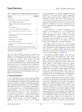
Table 1. Subtypes of non‑Hodgkin lymphomas in the spleen with immunoglobulin G and immunoglobulin M levels
recorded at 1,140 mg/dL and 47 mg/dL, respectively.
Subtypes References Tumor marker studies showed a slightly high soluble
B-cell type interleukin-2 receptor (666 U/mL, reference range: 122
α Splenic diffuse red pulp small B-cell lymphoma 1 – 496), whereas other tumor markers (CA19-9 and CEA,
α Splenic marginal zone lymphoma 1 determined by chemiluminescent enzyme immunoassay,
α Splenic B-cell lymphoma/leukemia with prominent 1 DUPAN-2, determined by enzyme immunoassay, and
nucleoli SPAN-1, determined by radioimmunoassay) were within
α Hairy cell leukemia 1 normal limits.
Primary splenic diffuse large B-cell lymphoma 2-10 Contrast-enhanced computed tomography (CT)
Fibrin-associated large B-cell lymphomas in splenic 11 revealed a mass measuring 5 cm × 4 cm with indistinct
cysts margins in the middle of the spleen (Figure 1A).
Primary splenic CD10-positive small B-cell lymphoma/ 12 18 F-fluorodeoxyglucose (FDG) positron emission
follicular lymphoma. tomography (PET)-CT confirmed the presence of an
T-cell and other cell type FDG-positive mass measuring 5 cm with a maximum
α Hepatosplenic T-cell lymphoma 1 standardized uptake value of 28.3 (Figure 1B). These
Splenic T/NK cell lymphoma 2 imaging studies suggested an intrasplenic lymphoma.
Splenic micronodular T-cell/histiocyte-rich large B-cell 13 The spleen (where the mass was initially thought
lymphoma to be located) and the tail of the pancreas were resected
Note: Refers to the World Health Organization Classification of en bloc through robot-assisted laparoscopic surgery, along
α
Tumors, 5 edition (Hematolymphoid tumors-Part B). Other splenic
1
th
lymphomas are based on literature surveys. with dissection of regional small lymph nodes. Gross
Abbreviation: NK: Natural killer. examination revealed a white, well-defined 5 cm soft mass
between the spleen and pancreas (Figure 1C).
type (46 cases), followed by splenic marginal zone Formalin-fixed paraffin-embedded tissue specimens
2
lymphoma (28 cases), and follicular lymphoma (11 cases). were stained with hematoxylin-eosin and specific
Shimono et al. classified primary splenic DLBCL into antibodies, including CD3 (Roche Diagnostics, United
3
two categories: (i) type A, which includes cases with States), CD5 (Roche Diagnostics, United States), CD10
or without lymphadenopathy of splenic hilum, and (Nichirei Biosciences, Japan), CD20 (Roche Diagnostics,
(ii) type B, characterized by cases with involvement of the United States), CD23 (Nichirei Biosciences, Japan),
bone marrow, liver, or peripheral blood, in addition to BCL-2 (Roche Diagnostics, United States), BCL-6 (Roche
the splenic lesions. Here, we report a rare case in which Diagnostics, United States), Ki-67 (Roche Diagnostics,
3
an intrasplenic mass was initially suspected based on United States), and MUM-1 (Roche Diagnostics, United
imaging studies but was later identified as DLBCL in the States), for immunohistochemistry (IHC). IHC was
splenic hilar lymph node without intrasplenic lesions after considered positive if more than 30% of tumor cells were
splenectomy examination.
positively stained. Ki-67 labeling index was estimated on
2. Case presentation the “hot spot” by simple visual inspection (“eyeballing”).
A 63-year-old Japanese man (174 cm, 64 kg), a hepatitis Microscopic examination of the resected mass revealed
B virus carrier, presented with a hypoechoic spleen mass that it did not invade the splenic parenchyma or pancreatic
discovered during a routine annual abdominal ultrasound. tissue, and no lymphoma cells were found in the regional
Notably, he reported no symptoms of left upper abdominal lymph nodes. Histopathological analysis confirmed the
pain or splenomegaly. His laboratory findings included a diagnosis of DLBCL, specifically of the germinal center
white blood cell count of 5,100 cells/µL, hemoglobin at B-cell type (Figure 1D-F). The bone marrow was free from
14.5 g/dL, platelet count of 250,000 platelets/µL, alanine lymphoma cell involvement, leading to a diagnosis of stage
aminotransferase at 14 U/L, lactate dehydrogenase at I DLBCL originating from the splenic hilar lymph node.
209 U/L (reference range: 124 – 222), total bilirubin of The karyotype was not assessed in this case. Fluorescence
®
™
0.58 mg/dL, total protein of 7.3 g/dL, and albumin at in situ hybridization (Vysis LSI IGH/MYC, CEP 8 Tri-
4.5 g/dL. Serological markers indicated active hepatitis color, Dual Fusion Translocation Probe, Abbott Molecular
B infection (HBsAg positive, HBsAb negative, HBcAb Inc., United States) analysis was performed on the
positive, HBeAb positive, and HCVAb negative). In lymphoma cells from the DLBCL tissue. Results indicated
addition, serum C-reactive protein was 0.1 mg/dL, negative for the IGH-MYC translocation, t(8;14)(q24;q32).
Volume 4 Issue 2 (2025) 101 doi: 10.36922/td.6742