
Page 110 - TD-4-2
P. 110
Tumor Discovery DLBCL in the splenic hilar lymph node
A B C
D E F
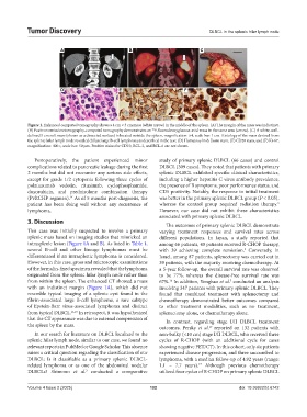
Figure 1. Enhanced computed tomography shows a 4 cm × 3 cm mass (white arrow) in the middle of the spleen. (A) The margin of the mass was indistinct.
(B) Positron emission tomography-computed tomography demonstrates an F-fluorodeoxyglucose-avid mass in the same area (arrow). (C) A white, well-
18
defined 5 cm soft mass (shown as a dissected surface) is located outside the spleen, magnification: ×4, scale bar: 1 cm. Histology of the mass derived from
the splenic hilar lymph node revealed diffuse large B-cell lymphoma as described in the text: (D) Hematoxylin & Eosin stain, (E) CD20 stain, and (F) Ki-67,
magnification: 400×, scale bar: 50 µm. Positive stains for CD10, BCL-2, and BCL-6 are not shown.
Postoperatively, the patient experienced minor study of primary splenic DLBCL (66 cases) and control
complications related to pancreatic leakage during the first DLBCL (309 cases). They noted that patients with primary
2 months but did not encounter any serious side effects, splenic DLBCL exhibited specific clinical characteristics,
except for grade 1/2 cytopenia following three cycles of including a higher hepatitis C virus antibody prevalence,
polatuzumab vedotin, rituximab, cyclophosphamide, the presence of B symptoms, poor performance status, and
doxorubicin, and prednisolone combination therapy CD5 positivity. Notably, the response to initial treatment
(PvRCHP regimen). As of 5 months post-diagnosis, the was better in the primary splenic DLBCL group (P < 0.05),
14
3
patient has been doing well without any recurrence of whereas the control group required radiation therapy.
lymphoma. However, our case did not exhibit these characteristics
associated with primary splenic DLBCL.
3. Discussion
The outcomes of primary splenic DLBCL demonstrate
This case was initially suspected to involve a primary varying treatment responses and survival rates across
splenic mass based on imaging studies that mimicked an different populations. In Japan, a study reported that
intrasplenic lesion (Figure 1A and B). As listed in Table 1, among 66 patients, 40 patients received R-CHOP therapy,
several B-cell and other lineage lymphomas must be with 39 achieving complete remission. Conversely, in
3
differentiated if an intrasplenic lymphoma is considered. Israel, among 87 patients, splenectomy was carried out in
However, in this case, gross and microscopic examinations 39 patients, with the majority receiving chemotherapy. At
of the formalin-fixed specimen revealed that the lymphoma a 5-year follow-up, the overall survival rate was observed
originated from the splenic hilar lymph node rather than to be 77%, whereas the disease-free survival rate was
from within the spleen. The enhanced CT showed a mass 67%. In addition, Yonghao et al. conducted an analysis
7
16
with an indistinct margin (Figure 1A), which did not involving 347 patients with primary splenic DLBCL. They
resemble typical imaging of a splenic cyst found in the found that combined treatment with splenectomy and
fibrin-associated large B-cell lymphoma, a rare subtype chemotherapy demonstrated better outcomes compared
of Epstein-Barr virus-associated lymphoma and distinct to other treatment modalities, such as no treatment,
from typical DLBCL. 11,15 In retrospect, it was hypothesized splenectomy alone, or chemotherapy alone.
that the CT appearance was due to external compression of In contrast, regarding stage I/II DLBCL treatment
the spleen by the mass. outcomes, Persky et al. reported on 132 patients with
17
In our search for literature on DLBCL localized to the non-bulky (<10 cm) stage I/II DLBCL, who received three
splenic hilar lymph node, similar to our case, we found no cycles of R-CHOP (with an additional cycle for cases
relevant reports in PubMed or Google Scholar. This absence showing negative PET/CT). In this cohort, only six patients
raises a critical question regarding the classification of our experienced disease progression, and three succumbed to
DLBCL: Is it classifiable as a primary splenic DLBCL- lymphoma, with a median follow-up of 4.92 years (range:
related lymphoma or as one of the abdominal nodular 1.1 – 7.7 years). Although previous chemotherapy
17
DLBCLs? Shimono et al. conducted a comparative utilized four cycles of R-CHOP on primary splenic DLBCL
3
Volume 4 Issue 2 (2025) 102 doi: 10.36922/td.6742