
Page 37 - TD-4-2
P. 37
Tumor Discovery Understanding glioblastoma invasion and therapy
7.3. Therapy resistance and other clinical growth-associated-protein 43 (GAP-43), and tweety-
consequences of GBM invasion homolog 1 (TTYH1). 136,137 Although they share many
Aggressive and diffuse invasion is a significant contributor features with other protrusive cellular structures, such as
to poor survival in GBM. Diffuse invasion creates tumors invadopodia and tunneling nanotubes (which are also found
138
with poorly defined margins that are impossible to resect in GBM), TMs are morphologically distinguished by their
136
completely with surgery. Early drastic attempts to remove remarkable length and capacity for long-term stability.
the entire affected brain hemisphere failed to prevent tumor Bona fide TMs are at least 50 µm long and have an average
2 136
recurrence on the contralateral side. Thus, while GBM cross-sectional area of approximately 1.5 µm . TMs have
123
136
initially presents as a distinguishable mass, diffuse invasion been observed to exceed 500 µm in vivo and commonly
fundamentally makes GBM a whole-brain disease. GBM surpass 1000 µm in in vitro models (Figure 5). TMs also
124
cells invading the blood vessels also strip the astrocytic exhibit significant plasticity in their temporal stability.
end feet from the endothelial basement membrane and They may be stable for weeks to months or dynamically
secrete enzymes that damage endothelial tight junctions remodeled to drive invasion at the tumor-brain interface. 136
and degrade the basal lamina. 82,125 8.2. TM networks
This invasive denuding and degrading of the TMs frequently arborize and interconnect into a
endothelium triggers reactive gliosis and disrupts the multicellular network. Cx43-mediated gap junctions are
neurovascular unit, thereby initiating a pathological evident at TM cross points and enable the TM network to
cascade that includes the loss of blood-brain barrier (BBB) function as a syncytium. TM networks bi-directionally
136
integrity, loss of activity-dependent blood flow regulation, propagate intercellular calcium waves (ICWs) similar to
serum leakage, and uncontrolled CNS access for ions, toxic those observed in the neurodevelopmental migration
and inflammatory molecules, and immune cells, impaired of neural progenitor cells 136,139 (Figure 6). TM networks
CNS uptake of glucose and oxygen, hypoxia, necrosis, and also exchange signaling molecules and traffic
140
edema. 82,125,126 organelles, including mitochondria. TM length and
136
In contrast, intraparenchymal invasion carries cells far quantity increase with increasing astrocytoma grade,
beyond the margin of the radiation target and into areas of but they are not regularly observed in 1p/19q co-deleted
136
the brain with a robustly intact BBB, where they are largely oligodendrogliomas, which may partially explain the
inaccessible to immune cells and therapeutic drugs. Some therapeutic response and survival difference between the
127
evidence additionally suggests that invasive astrocytes astrocytoma and oligodendroglioma cohorts.
may limit cell cycle progression and could, therefore, be
less sensitive to conventional therapies that generally 8.3. Molecular understanding of TMs
target proliferating cells. 128-130 Proteolysis and glutamate- Molecular drivers of TM formation have thus far been
mediated intraparenchymal invasion also physically erode predominantly identified through in silico comparison
the normal neural architecture, thereby disrupting circuit
control, triggering seizures, and ultimately leading to
functional deterioration. 131,132
In addition, many glioma therapies are known to
exacerbate the invasive motility that drives tumor recurrence
and neurological decline. 49,50,133,134 Most GBMs exhibit
invasive behavior and significant therapy resistance at the
time of tumor recurrence. Thus, invasive motility remains
a primary obstacle to the successful treatment of GBM. 135
8. GBM TMs
8.1. TM structure and morphology
TMs are invasive neurite-like protrusions that extend
from the cell bodies of diffuse astrocytoma cells into the
surrounding brain parenchyma. TMs are structurally
136
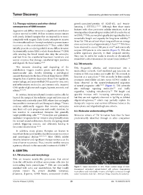
enriched with actin and microtubules, but they locally Figure 5. Tumor microtubes in vitro. Phase contrast image of three-
dimensional networked patient-derived cell cultures demonstrates
express myosin IIa, protein disulfide isomerase, neurospheres and individually invasive cells highly connected through
β-catenin, β-parvin, GFAP, Nestin, connexin43 (Cx43), tumor microtubes. Scale bar = 1000 µm. Image created by the author(s).
Volume 4 Issue 2 (2025) 29 doi: 10.36922/td.8578