
Page 89 - AIH-1-3
P. 89
Artificial Intelligence in Health Dental cavity prediction with computer vision
of bitewing images in clearly capturing such lesions. heralding a new era of precision and efficiency in detecting
19
Another innovative approach involved classifying tooth dental caries. As we delve further into this article, we
caries using quantitative light-induced fluorescence (QLF) will explore the mechanics behind these innovations,
images with the help of the Xception deep learning model, their practical applications, and the challenges and
underscoring the significance of image augmentation future directions in integrating advanced computational
and K-fold cross-validation in training robust models. techniques into dental care.
20
A systematic review aimed at evaluating neural networks
in caries detection highlighted the diverse methodologies 3. Methods
and neural network architectures employed across studies, 3.1. Data collection and pre-processing
reflecting the dynamic evolution of AI applications in
dental diagnostics. 21 The image data were sourced from Kaggle (https://
www.kaggle.com/datasets/salmansajid05/oral-
Further illustrating the potential of machine learning diseases?resource=download-directory). This dataset
in dentistry, a previous study applied several algorithms, comprises a collection of images obtained from multiple
notably random forest, achieving high performance in health centers and reliable dental websites, ensuring the
predicting the risk of dental caries from a dataset derived variety and validity of the dental conditions depicted. Each
22
from a children’s oral health survey. A systematic review image in the dataset is thoroughly marked with bounding
focusing on AI for radiographic imaging detection of boxes, accurately representing the dental condition.
caries lesions critically evaluated studies, revealing a
preference for CNN models in most research, with a range 3.1.1. Description of the colored image dataset
from 15 to 2900 radiographs used across various studies The colored image dataset used in this study comprises a
to build AI models. The use of deep learning for caries total of 218 dental cavity images captured using a standard
23
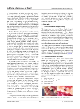
detection through tooth surface segmentation in intraoral device camera. These images were obtained under casual
photographic images has been investigated, employing conditions, featuring open jaws and clear representations
U-Net for segmentation and ResNet-18 and Faster R-CNN of dental cavities. The dataset served as the foundational
for classification and localization, thereby reducing false source of visual data for training and evaluation (Figure 1).
alarms and enhancing detection accuracy. Another study
24
developed a CNN model for diagnosing dental caries from 3.1.2. Data augmentation techniques
bitewing radiographs, demonstrating the utility of deep
learning in enhancing dental diagnostic processes. 19 Data augmentation plays a pivotal role in expanding the
dataset and enhancing model robustness. To achieve
A research endeavor introduced a novel method for this, we leveraged the Image Data Generator, an image
classifying dental caries using QLF imaging combined augmentation API integrated within Keras – an open-source
with CNNs, aiming to improve accuracy in real-time Python library for machine learning. ImageDataGenerator
caries detection in clinical settings. Lian et al., utilized enabled artificially diversifying the dataset by applying
25
20
deep learning methods to detect and classify caries lesions transformations such as rotation, shifting, zooming,
on panoramic films, comparing performance with expert shearing, and reflection. These augmentations fostered
dentists and showing similar accuracy and reliability. the development of more adept models and improved
Alharbi et al. applied nested U-Net models to dental their ability to generalize across various scenarios. In our
26
panoramic X-ray images for caries detection, demonstrating experimentation, the augmentation parameters were set as
high testing accuracy and robust model performance. follows:
Sikri et al., presented a comprehensive narrative i. Rotation range: 40°
27
review on the applications of AI in dentistry, detailing ii. Width and shifting range: 0.2
how AI integrates into various aspects of dental care, iii. Zoom range: 0.2
from diagnostics to patient management. Meanwhile, iv. Shear range: 0.2.
Zhou et al. explore a more focused application with
28
their development of a context-aware CNN specifically
designed for diagnosing caries in children from dental
panoramic radiographs, demonstrating the potential of
machine learning to address unique challenges in pediatric
dentistry.
These studies collectively underscore the transformative
impact of machine learning and AI on dental diagnostics, Figure 1. Colored image with a single cavity and multiple cavities
Volume 1 Issue 3 (2024) 83 doi: 10.36922/aih.3184