
Page 73 - AIH-2-2
P. 73
Artificial Intelligence in Health Improved liver tumor segmentation with dense networks
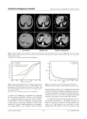
Figure 5. Selected examples of pre-processed CT images for the three different pre-processing methods: HU value clipping with a preset CT window
(Window Clip), the full range of HU values without any window (No Window), and the proposed integrated window optimization pre-processing module
(Window Optimization).
Abbreviations: CT: computed tomography; HU: Hounsfield unit.
Figure 6. Input–output mapping curves of the different pre-processing Figure 7. Training loss curves of 2D DenseUNet and I -DenseFCN
2
methods: HU value clipping with a preset CT window (Window Clip), Abbreviation: I -DenseFCN: intra- and inter-block densely connected FCN.
2
the full range of HU values without any window (No Window), and
the proposed integrated window optimization pre-processing module network, whereas Chlebus et al. employed an object-level
41
(Window Optimization).
Abbreviations: CT: computed tomography; HU: Hounsfield unit. random forest classifier. Despite not utilizing extensive post-
processing operations, the proposed method achieved a
to improve the segmentation performance by enhancing Dice per case score of 71.3% for lesion segmentation through
the representation capabilities of features, as demonstrated parameter tuning, hence outperforming other methods.
by a consistent performance gain from UNet , ResNet Our approach exceeds the baseline methods from the
39
30
to DenseUNet in Table 4. In addition, other approaches International Symposium on Biomedical Imaging and
29
leverage some post-processing techniques to filter out false MICCAI challenges according to the leaderboard listed
positives for improved tumor segmentation performance. in Bilic et al. and surpasses the current best 2D-based
36
For example, Bellver et al. trained a lesion detection method DenseUNet (Table 4) on the LiTS dataset. It also
40
Volume 2 Issue 2 (2025) 67 doi: 10.36922/aih.5001