
Page 135 - AIH-2-3
P. 135
Artificial Intelligence in Health Early detection of CIN/cervicitis using ML
ratio values indicate normal tissues, while higher ratio weakness, and loss of appetite. The physical examination
values represent higher grades of malignancy. The most showed an irregular mass on the anterior side of the cervix,
malignant site was identified as a site with the highest DR with the involvement of the posterior and lateral fornix,
image ratio R610/R545 value, and a biopsy was obtained as well as the lower part of the vagina. The histopathology
from this site for an accurate determination of the cancer result following cervical biopsy confirmed malignancy.
grade through histopathology. Immunohistochemistry and immunofluorescence, along
Typical cases of patients with cervical intraepithelial with histopathology results, confirmed the diagnosis of
neoplasia (CIN) and one case of cervicitis are presented carcinosarcoma (squamous cell carcinoma with sarcoma),
below: with an R610/R545 image ratio value of 3.005, as illustrated
in Figure 4.
3.1.1. Case 1
3.1.3. Case 3
Figure 3 shows a 56-year-old patient with complaints
of post-menopausal bleeding and abdominal pain. The A 50-year-old patient with cervicitis was studied based
gynecologist identified a massive growth covering the on the DR image ratio R610/R575 value. The patient had
anterior and posterior lips of the cervix and the upper part difficulty in passing urine, and her cervix showed erosions
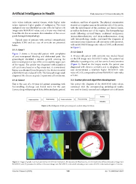
of the vagina. The patient was diagnosed with squamous (Figure 5). Based on the biopsy results, the patient was
cell carcinoma based on a Pap smear test. A conventional diagnosed with chronic cervicitis and no dysplasia. This
biopsy was obtained from the growth. Our device recorded case demonstrated a relatively higher R610/R575 ratio
a R610/R545 ratio value of 2.078. The histopathology result value of 2.433, compared to a lower R610/R545 ratio value
diagnosed the disease as grade 2 squamous cell carcinoma. of 1.461.
3.1.2. Case 2 3.2. Scatter plot and algorithm development
This is the case of a 50-year-old patient presenting with The scatter plot diagram of the R610/R545 ratio values,
foul-smelling discharge and blood stains for the past correlated with the corresponding pathological results,
2 years, along with passage of clots, abdominal pain, general was used to classify normal and malignant cervical tissues
Figure 2. Block diagram of data processing and analysis using CerviScan
A B C
D
Figure 3. Images of grade 2 cervical carcinoma: (A) Clinical impression, (B) pseudocolor fluorescence, (C) monochrome diffuse reflectance image ratio
R610/R545, and (D) pseudocolor diffuse reflectance image ratio R610/R545, with a dial indicating a ratio value of 2.078 in the marked region of interest
Volume 2 Issue 3 (2025) 129 doi: 10.36922/aih.8527