
Page 133 - AIH-2-3
P. 133
Artificial Intelligence in Health Early detection of CIN/cervicitis using ML
gingiva using the R620/R575 image intensity ratio. In cervicitis, respectively. The study conducted using this
another study, the R620/R575 spectral ratio was able to device demonstrates its potential for real-time tissue status
discriminate between healthy gingiva from gingivitis, with assessment and its ability to detect the most malignant site
90% sensitivity and 94% specificity, whereas a sensitivity for biopsy. The DR image ratios were correlated with the
of 91% and a specificity of 100% were obtained when histopathology results of guided biopsies to develop an ML
discriminating gingivitis from periodontitis. Recently, algorithm. The diagnostic accuracy of the screening was
18
Barik et al. recorded the fluorescence emission of the determined from the scatter plot diagrams, and the results
19
cervix on excitation at 325 nm, and the spectral features were presented accordingly.
were used to discriminate dysplasia from inflammatory
changes. An increase in fluorescence emission intensity 2. Materials and methods
in cervicitis samples was observed at 435 nm compared 2.1. Methodology
to normal tissue, which was attributed to the fluorophore
nicotinamide adenine dinucleotide. Zhang et al. utilized a The CerviScan (Figure 1) consisted of a bimodal imaging
20
feature fusion method to extract information from Raman camera designed for fluorescence and DR imaging of
spectra and its derivatives. They classified inflamed cervical the cervix. The device was equipped with a 5-megapixel
tissues as high-grade squamous intraepithelial lesions and monochrome camera (MU9PM-MH, Ximea, GmbH,
LSIL based on the intensity of the prominent spectral Germany) to record the fluorescence emission of collagen
−1
peaks at 548, 640, 1,452, and 1,664 cm . In another study, on excitation with 370 nm LEDs (ATS2012UV365,
colposcopy images, in conjunction with deep learning, Kingbright, United States). The HbO absorption changes
2
achieved an accuracy of 95.2% in discriminating chronic in cervical tissues were assessed from the DR images
cervicitis from cervical cancer. 21 captured under illumination with LEDs emitting at
545 nm (L1C1-GRN1000000, LUMI LEDs, Netherlands)
A recent study combining fluorescence and DR imaging and 575 nm (SMP2-SGC, Bivar, United States), with both
for the detection of oral cavity lesions reported improved overlapping the HbO absorption spectra at 542 nm and
2
diagnostic accuracy in discriminating potentially malignant 577 nm. The DR images were also captured following
oral lesions from normal tissues. This was achieved using a LED illumination at 610 nm (SMP2-SOC, Bivar, United
machine learning (ML) algorithm based on the DR image States), where the absorption changes in cervical tissues
ratio R620/R545, which represents changes in the deoxy to due to Hb were stronger compared to HbO . A Windows
2
oxygenated Hb absorption in tissue. The study highlights tablet (Chuwi, China) was connected to a USB camera
8
the effectiveness of non-invasive screening modalities (Ximea, GmbH, Germany), with proprietary software
to provide real-time user feedback and to enhance installed for camera control, image capture, and analytics.
compliance, particularly for the early detection of cervical The monochrome images (Im545, Im610, Im575, and
cancers. F370) recorded were processed by software in real time
In the past decade, ML models have been widely used to generate ratio images (R610/R545 and R610/R575)
for medical diagnosis. Dong et al. developed an ML model and their corresponding pseudocolor maps (PCM). The
22
for cervical cancer risk stratification using full-genotyping region of interest (ROI) was then marked on these ratio
of high-risk HPV test data. The study compared four ML
models zs– XGBoost, support vector machine (SVM),
random forest, and naïve Bayes – where the XGBoost
model was found to be the most effective model.
Surface plasmon resonance biosensor with ML
optimization was developed by Wekalao et al. for cervical
23
cancer detection. This support vector regression was found
to enhance the sensor’s predictive capability, reducing the
stimulation time by 80%. Several deep learning techniques
have been used for cervical cancer diagnosis using
pathology slides and colposcopy images. 24
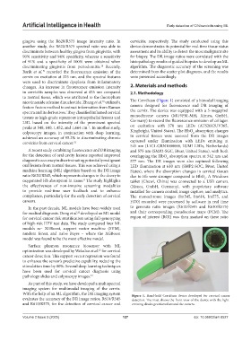
As part of this study, we have developed a multispectral
imaging system for multimodal imaging of the cervix.
With the help of an ML algorithm, the DR imaging system Figure 1. Hand-held CerviScan device developed for cervical cancer
evaluates the accuracy of the DR image ratios, R610/R545 detection. The inset shows the front view of the device with the light-
and R610/R575, for the detection of cervical cancer and emitting diodes positioned around the camera.
Volume 2 Issue 3 (2025) 127 doi: 10.36922/aih.8527