
Page 138 - AIH-2-4
P. 138
Artificial Intelligence in Health Artificial intelligence app for EVD navigation
A B C D
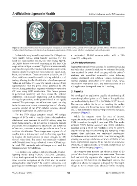
Figure 2. Schematic representation of neuronavigation using an iOS mobile device. (A) Anatomic review and target selection. (B) An iOS device mounted
with the patient’s head in view. (C) Evaluation of registration parameters. (D) Procedural interface for alignment and depth guidance.
Industrial-strength models, optimized for iOS systems, a randomized 10-fold cross-validation with a 90%
were rapidly trained using transfer learning. For the train/10% testing split.
head CT segmentation model, the open-source QURE.
AI CQ500 dataset was used, consisting of 491 head CTs 2.4. Model performance
acquired on multiple scanners. Eight scans were manually Segmentation models were assessed for accuracy in training
45
annotated to segment key anatomical structures relevant to and validation cohorts. In addition, we evaluated the initial
trauma surgery: background, extracranial soft tissue, skull, time required for point cloud merging with the patient’s
brain, and ventricles. These annotations yielded 4,096 CT anatomy and quantified cumulative error following
slices, which were used for model training, validation, and scaling, alignment, and rotation. Finally, performance
testing, allowing for the identification of each component metrics included intersection over union (I/U), varied
within an individual CT scan. Our model combines these intersection over union (I/U), and inference times of the
segmentations with the point cloud generated by iOS iOS application during real-time EVD tracking.
devices, fusing point cloud segments with the pre-operative
CT scan using GPU acceleration. This fusion process 3. Results
is performed iteratively and then creates the optimal
alignment, continuously registering and re-registering We developed an application capable of performing all
steps of surgical navigation on iOS devices. The application
during the procedure, as the patient’s head is not rigidly
secured. The system operates without user input, ensuring can load and display head CTs in DICOM or NIFTI format.
instantaneous, continuous neuronavigation and allowing The surgeon selects the target by touching the mobile
accurate overlay of the EVD catheter location without device’s screen, and the device stores that information for
requiring head fixation or a reference array. the 3D transformations necessary to perform the navigated
procedure (Figure 3).
For the EVD object detection model, 937 unique
images of EVDs with a visually distinct dodecahedron While the surgeon views the scan of interest,
attachment were acquired in an ICU setting using the segmentation is performed in the background by a UNet
front-facing camera of an iOS device to simulate bedside CNN trained on the eight 1 mm head CTs. This model
placement. The dodecahedron, attached to the distal end achieved 98.3% testing accuracy and 98.2% validation
of the EVD, contained a unique QR code on each face to accuracy using a 50/50 test–validation split. To confirm
facilitate identification. These images were segmented and that the model was not overfitting and remained robust
used to train a feature-based machine learning algorithm against class imbalance, we performed randomized
to localize the dodecahedron in space through the iOS 10-fold cross-validation with a 90% train/10% testing split,
device’s video feed. The model was externally validated yielding an average validation accuracy of 98.3% across
with 200 additional images obtained in non-ICU settings. folds. Segmentation requires 30 ms per slice on a standard
In total, 700 randomly selected images were used for iPhone 12, or approximately 3 s per scan, and provides the
training and 237 for validation. data for surface merging (Figure 4).
Two models were trained: a full network and a transfer- The surgeon then mounts the phone in front of the
learning model using YOLOv2 (YOLOv2, Ultralytics, patient’s head and navigates to the next screen. The video
USA), both optimized for iOS systems to achieve real- feed semantically segments the largest head in view
time, accurate navigation of the EVD and its custom and captures 3D data from the TrueDepth camera. The
dodecahedron attachment. To confirm robustness and application allows the surgeon to inspect the TrueDepth
49
guard against overfitting or class imbalance, we performed image in 3D to ensure scan adequacy before merging.
Volume 2 Issue 4 (2025) 132 doi: 10.36922/aih.8195