
Page 20 - AN-1-2
P. 20
Advanced Neurology MiR-195 regulates MS-dCA1 neural circuit in CBH rat
A B C
D E
F G H I
J K L
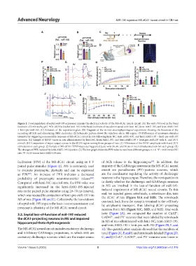
Figure 1. Downregulation of septal miR-195 expression impairs the electrical activity of the MS-dCA1 neural circuit. (A) The miR-195 level in the basal
forebrain of 2VO rats by qRT-PCR. (B) The level of miR-195 in the basal forebrain of rats administered with lenti-NC, lenti-AMO-195, and lenti-AMO-195
+ lenti-pre-miR-195. (C) Schedule of the experiment plan. (D) Diagram of the in vivo electrophysiological experiment showing the locations of the
recording (dCA1) and stimulating (MS) electrodes. (E) Schematic picture shows the injection site in MS region. (F) Differences of minimum stimulus
intensity for triggering a measurable response of MS-dCA1 circuit in rats following lenti-NC, lenti-AMO-195, and lenti-AMO-195 + lenti-pre-miR-195
injection. (G) Sample of fEPSP traces in rats administrated by lenti-NC, lenti-AMO-195, and lenti-AMO-195 + lenti-pre-miR-195 with 8- and 20-V
stimuli. (H) Comparison of input-output curves in the dCA1 region among three groups of rats. (I) Differences of the fEPSP amplitude with 8 and 20 V
stimulation in each group. (J) Sample of MS-dCA1 fEPSP traces during paired pulse with 20, 40, and 70 ms of interstimulus intervals for each group. (K)
The changes of PPR induced by lenti-AMO-195 injection. (L) The bar graph shows the PPR value in rats from different groups. n = 6. *P < 0.05 versus NC
rats. P < 0.05 versus lenti-AMO-195 rats.
#
facilitation (PPF) of the MS-dCA1 circuit using an 8 V of ACh release in the hippocampus . In addition, the
[35]
paired-pulse stimulus (Figure 1J). PPF is commonly used majority of the GABAergic neurons in the MS-dCA1 neural
to evaluate presynaptic plasticity and can be expressed circuit are parvalbumin (PV)-positive neurons, which
as PPR . An increase of PPR indicates a decreased are the coordinator regulating the activity of cholinergic
[33]
probability of presynaptic neurotransmitter release . neurons in the hippocampus. Therefore, the next question is
[34]
Compared with lenti-NC-injected rats, the PPR value was to clarify whether the cholinergic and GABAergic neurons
significantly increased in the lenti-AMO-195-injected in MS are involved in the loss-of-function of miR-195-
rats under paired-pulse stimulus using 20–70 ms interval, induced impairment of MS-dCA1 neural circuits. To this
which was rescued by coinjection of lenti-pre-miR-195 into end, we injected green retrobeads, a retrotracer dye, into
MS of rats (Figure 1K and L). Collectively, the knockdown the dCA1 of rats (Figure S1A and S1B). The retrobeads
can track back from the synaptic terminal to the cell body
of septal miR-195 impairs the basic neurotransmission and by axoplasmic transport, thus labeling dCA1-projecting
presynaptic plasticity of the MS-dCA1 neural circuit. neurons from MS (Figure S1C, S1D, and S1E). One week
+
3.2. Septal loss-of-function of miR-195 reduced later (Figure 2A), we compared the number of ChAT ,
+
+
the dCA1-projecting neurons in MS and impaired GAD67 , and PV neurons that were labeled by retrobeads
hippocampal theta rhythmogenesis in MS of rats administered with lenti-NC, lenti-AMO-195,
and lenti-AMO-195 + lenti-pre-miR-195 (Figure 2B, E, and
The MS-dCA1 neural circuit includes excitatory cholinergic H). The quantification analysis showed that the numbers of
and inhibitory GABAergic projections, in which 66% are total (Figure 2C, F, and I) and retrobeads-labeled (Figure 2D,
excitatory cholinergic neurons which are the major source G, and J) ChAT , GAD67 , and PV neurons were lower in
+
+
+
Volume 1 Issue 2 (2022) 5 https://doi.org/10.36922/an.v1i2.116