
Page 11 - AN-1-3
P. 11
Advanced Neurology Long-term in vivo MRI tracking of SPIO-labeled NSCs
A B
C D
E
F
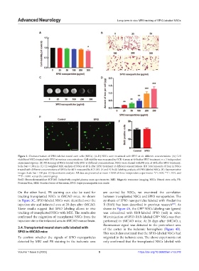
Figure 1. Characterization of SPIO-labeled neural stem cells (NSCs). (A–D) NSCs were incubated with SPIO at six different concentrations. (A) Cell
viability of NSCs treated with SPIO at various concentrations. Cell viability was measured by CCK-8 assay at 48 h after SPIO treatment. n = 3 independent
experiments/group. (B) PB staining of NSCs treated with SPIO at different concentrations. NSCs were stained with PB iron at 48 h after SPIO treatment.
Scale bar = 100 μm. (C) T2-weighted MRI analysis of NSCs at 48 h after SPIO treatment at different concentrations. (D) Total amounts of iron in NSCs
treated with different concentrations of SPIOs for 48 h measured by ICP-MS. (E and F) BrdU labeling analysis of SPIO-labeled NSCs. (E) Representative
images. Scale bar = 100 μm. (F) Quantitative analysis. All data are presented as mean ± SEM of three independent experiments. *P < 0.05, **P < 0.01, and
***P < 0.001 versus the control group.
BrdU: Bromodeoxyuridine: ICP-MS: Inductively coupled plasma mass spectrometer, MRI: Magnetic resonance imaging, NSCs: Neural stem cells, PB:
Prussian blue, SEM: Standard error of the mean, SPIO: Superparamagnetic iron oxide.
On the other hand, PB staining can also be used for are carried by NSCs, we examined the correlation
tracking transplanted NSCs in tMCAO mice. As shown between transplanted NSCs and SPIO nanoparticles. The
in Figure 3C, SPIO-labeled NSCs were identified over the synthesis of SPIO nanoparticles labeled with rhodamine
injection site and infarcted area at 28 days after tMCAO. B (RhB) has been described in previous research . As
[25]
These results suggest that SPIO labeling allows in vivo shown in Figure 4A, the GFP NSCs labeling rate (green)
+
tracking of transplanted NSCs with MRI. The results also was colocalized with RhB-labeled SPIO (red) in vitro.
confirmed the migration of transplanted NSCs from the Microinjection of SPIO-RhB-labeled GFP NSCs was then
+
injection site to the infarcted area in tMCAO mouse brain. performed in tMCAO mice. At 28 days after tMCAO, a
fluorescence signal was detected in the peri-infarct area
3.4. Transplanted neural stem cells labeled with of the cortex in the ischemic hemisphere (Figure 4B).
SPIO in tMCAO mice This result demonstrated that the SPIO-labeled NSCs had
To confirm whether the signals of SPIO nanoparticles migrated to the ischemic area. The above experiments not
detected by MRI and PB staining in the ischemic area only confirmed that the transplanted NSCs labeled with
Volume 1 Issue 3 (2022) 5 https://doi.org/10.36922/an.v1i3.278