
Page 12 - AN-1-3
P. 12
Advanced Neurology Long-term in vivo MRI tracking of SPIO-labeled NSCs
A B
C D
E
F
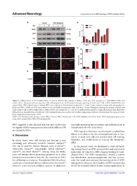
Figure 2. Differentiation of SPIO-labeled NSCs. (A and B) Western blot analysis of Nestin, GFAP, and TUJ1 expression in SPIO-labeled NSCs and
control-NSCs. Timescale indicates days after NSC differentiation. (C–F) Immunofluorescence staining of GFAP and TUJ1 in SPIO-labeled NSCs and
control-NSCs. NSCs labeled with or without SPIO were cultured in differentiation medium for 1, 7, and 14 days, and then stained with cells positive for
GFAP and TUJ1. GFAP and TUJ1 were stained in red and DAPI was stained in blue. Scale bar = 50 μm. Histogram showing the number of GFAP and
TUJ1-positive cells in NSCs labeled with or without SPIO and cultured for 1, 7, and 14 d in differentiation medium. Timescale indicates days after NSCs
differentiation. Quantification of GFAP and TUJ1 immunofluorescence-positive cell numbers using ImageJ software. Data represent the mean of at least
three independent experiments ± SEM.
GFAP: Glial fibrillary acidic protein, control-NSCs: Control, NSCs: Neural stem cells, SEM: Standard error of the mean, SPIO: Superparamagnetic iron
oxide, SPIO-labeled NSCs: SPIO, TUJ: β-tubulin III.
SPIO migrated to the infarcted side, but also verified that especially pertaining the persistence and differentiation of
the signals of SPIO nanoparticles detected by MRI and PB transplanted cells into brain tissue.
are carried by NSCs. With regard to this issue, we developed a method that
4. Discussion allows us to observe the fate of transplanted cells in vivo,
that is, to track stem cells and monitor stem cell homing,
In recent years, stem cell therapy has become a very migration, and proliferation to evaluate the therapeutic
promising and advanced scientific research method effect.
[26]
that can be used for various diseases, such as stroke , In the present work, we developed a novel cell label-
[27]
Parkinson’s disease , amyotrophic lateral sclerosis , ing strategy based on SPIO nanoparticles and explored its
[29]
[28]
[31]
cancer , and heart disease . Among them, for stroke, application for long-term MRI imaging tracking of trans-
[30]
stem cell therapy shows promising prospects as it offers planted NSCs. The SPIO nanoparticles showed a narrow
potential neurorestorative benefits. The potential of NSCs size distribution, superparamagnetism, and low cytotox-
for cell therapy is enormous. Transplanted NSCs have been icity. Our result demonstrated the tracking ability of SPIO
used in pre-clinical and clinical trials to restore function . nanoparticles. However, the biosafety of SPIO nanoparticles
[32]
However, some relevant problems need to be addressed, should be evaluated before further clinical application. In
Volume 1 Issue 3 (2022) 6 https://doi.org/10.36922/an.v1i3.278