
Page 49 - AN-2-3
P. 49
Advanced Neurology TRPM7 signaling in glioblastoma
After 24 h, the invaded cells were fixed with methanol TRPM7, either directly or indirectly, thus indicating that
and stained using 1% Toluidine blue. Four fields of each both proteins potentially participate in the same signaling
chamber were imaged using a phase-contrast Olympus pathway to regulate GBM cell function.
microscope (×10 objective) and the cells were quantified
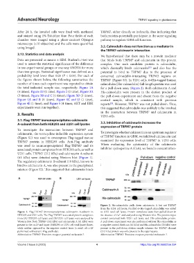
using ImageJ. 3.2. Calmodulin does not function as a mediator in
the TRPM7-calcineurin interaction
2.12. Statistics and data analysis We hypothesized that there may be a protein mediator
Data are presented as means ± SEM. Student’s t-test was that binds both TRPM7 and calcineurin in this protein
used to assess the statistical significance of the difference complex. One such candidate protein is calmodulin,
in two experimental groups or one-way ANOVA for more which classically binds calcineurin and also has the
[25]
than two groups. Statistical significance was defined as a potential to bind to TRPM7 due to the presence of
probability level lower than 0.05 (P < 0.05). For each of conserved calmodulin-interacting TRPM3 regions on
the figures shown below, the following summarizes the TRPM7 (Figure S3). In U251 cells, 6×His-tagged human
number of times each experiment was repeated to obtain calmodulin (His-calmodulin) full-length protein was used
the total indicated sample size, respectively: Figure 1A for a pull-down assay (Figure 2). Both calcineurin A and
(4 times), Figure 1B (1 time), Figure 2 (1 time), Figure 3A His-calmodulin were present in the elution product of
(3 times), Figure 3B and C (4 times), Figure 3D (3 times), the pull-down experiment and absent from the negative
Figure 4A and B (6 times), Figure 4C and D (1 time), control sample, which is consistent with previous
Figure 4E (1 time), and Figure 5 (4 times; AKT and ERK reports . However, TRPM7 was not pulled down. Thus,
[25]
experiments were run together). this suggested that calmodulin was unlikely to be involved
in the interaction between TRPM7 and calcineurin in
3. Results U251 cells.
3.1. Flag-TRPM7 immunoprecipitates calcineurin
A-subunit from both HEK293 and U251 cell lysates 3.3. Inhibition of calcineurin increases the
expression of TRPM7 protein
To investigate the interaction between TRPM7 and
calcineurin, the tetracycline-inducible expression system To investigate whether calcineurin is an upstream regulator
(Figure S1) was used to overexpress Flag-tagged mouse of TRPM7 function in GBM, we inhibited calcineurin and
TRPM7 protein in HEK293 cells. Anti-Flag antibody examined the expression level of TRPM7 in U251 cells.
was used to immunoprecipitated Flag-TRPM7 and its When evaluating the cytotoxicity of the calcineurin
associated protein complexes from HEK293 cells, as well as inhibitor cyclosporine A (CsA), we found a concentration-
U251 cells. TRPM7 (212 kDa) and calcineurin A-subunit
(61 kDa) were detected using Western blot (Figure 1).
The regulatory calcineurin B-subunit (19 kDa), known to
bind to calcineurin A, was also present in the precipitated
mixture (Figure S2). This suggested that calcineurin binds
A B
Figure 2. His-calmodulin pulls down calcineurin A but not TRPM7
from the U251 cell lysate. Purified 6×His-tagged calmodulin was added
Figure 1. Flag-TRPM7 immunoprecipitates calcineurin A-subunit in to U251 total cell lysate. Protein complexes were then pulled down in
HEK293 and U251 cells. The Flag-TRPM7-associated protein complexes the absence of Ca and analyzed using Western blot. The positive input
2+
from (A) HEK293 cell lysate and (B) U251 cell lysate were analyzed by control contained both U251 cell lysate and His-calmodulin protein.
Western blot. Both TRPM7 (212 kDa) and calcineurin A (61 kDa) were A pull-down experiment was also performed without His-calmodulin as
present in the co-IP and input (HEK293 or U251 cell total lysate) lanes, a negative control. Both can (61 kDa) and His-calmodulin (20 kDa) were
while neither appeared in the negative control lanes (a result of co-IP present in the pull-down elution sample whereas the TRPM7 channel
performed without anti-Flag antibody). (212 kDa) protein was only present in the supernatants.
Abbreviation: TRPM7: Transient receptor potential melastatin 7. Abbreviation: TRPM7: Transient receptor potential melastatin 7.
Volume 2 Issue 3 (2023) 4 https://doi.org/10.36922/an.334