
Page 51 - AN-2-3
P. 51
Advanced Neurology TRPM7 signaling in glioblastoma
A C
B
D
E
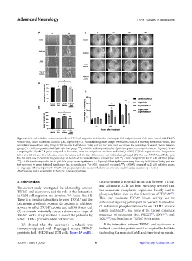
Figure 4. CsA and naltriben co-treatment reduces U251 cell migration and invasion similarly to CsA-only treatment. Cells were treated with DMSO
vehicle, CsA, and/or naltriben (10 and 25 µM, respectively). (A) Wound healing assay. Images were taken 0 and 24 h following the scratch wound, and
wound size was outlined using ImageJ. (B) One-way ANOVA and Tukey post hoc test were used to compare the percentage of wound closure between
groups (*p < 0.05; compared to the 25 µM naltriben group: #### p < 0.0001; and compared to the 10 µM CsA group: ns, no significance; n = 7/group). When
comparing the 10 µM CsA group compared to the control, there was a significant moderate reduction (p < 0.05). (C) Oris migration assay. Images were
taken at 0, 12, 24, and 48 h following wound formation, and the size of the wound was outlined using ImageJ. (D) One-way ANOVA and Tukey post
hoc test were used to compare the percentage of closure of the wound between groups (*p < 0.05; **p < 0.01; compared to the 25 µM naltriben group:
#### p < 0.0001; and compared to the 10 µM CsA group: ns, no significance; n = 7/group). € Matrigel invasion assay. One-way ANOVA and Tukey post hoc
test were used to assess statistical significance (ns, no significance; **p < 0.01 compared to control; #### p < 0.0001 compared to 25 µM naltriben group;
n = 3/group). When comparing the 10 µM CsA group compared to the control, there was a trend toward moderate reduction (p = 0.131).
Abbreviations: CsA: Cyclosporine A; ANOVA: Analysis of variance.
4. Discussion thus suggesting a potential interaction between TRPM7
and calcineurin A. It has been previously reported that
The current study investigated the relationship between
TRPM7 and calcineurin, and the role of this interaction the calcineurin phosphatase region can directly bind to
[27]
in GBM cell migration and invasion. We found that: (1) phosphorylation sites on the C-terminus of TRPM7 .
There is a possible interaction between TRPM7 and the This may modulate TRPM7 kinase activity and its
[28]
calcineurin A-subunit proteins; (2) calcineurin inhibition subsequent signaling pathways . In contrast, the function
appears to affect TRPM7 protein and mRNA levels; and of N-terminal phosphorylation sites on TRPM7 remains
[29]
(3) calcineurin potentially acts as a downstream target of largely undefined , and none of the known consensus
[31]
[30]
TRPM7 and is likely involved in one of the pathways by sequences of calcineurin (i.e., PXIXIT , LXVP , and
which TRPM7 promotes GBM cell function. LQLP ) are found at the TRPM7 N-terminus.
[32]
We showed that the calcineurin A-subunit was If the interaction between TRPM7 and calcineurin is
immunoprecipitated with Flag-tagged mouse TRPM7 indirect, a mediator protein would be required to facilitate
protein in both HEK293 and U251 cells (Figure 1A and B), the binding. Calmodulin (CaM), a calcium-binding protein,
Volume 2 Issue 3 (2023) 6 https://doi.org/10.36922/an.334