
Page 134 - AN-3-3
P. 134
Advanced Neurology Delayed occlusion after mechanical thrombectomy
A B C D
E
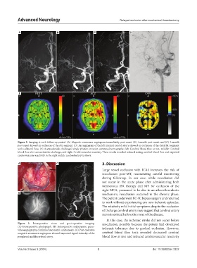
Figure 2. Imaging at each follow-up period. (A) Magnetic resonance angiogram immediately post-onset, (B) 1-month post-onset, and (C) 3-month
post-onset showed an occlusion of the M1 segment. (D) An angiogram of the left internal carotid artery showed an occlusion of the distal M1 segment
with collateral flow. (E) Acetazolamide-challenged single photon emission computed tomography; left: Cerebral blood flow at rest, middle: Cerebral
blood flow after acetazolamide challenge, and right: Cerebrovascular reactivity. These results revealed reduced resting cerebral blood flow and impaired
cerebrovascular reactivity in the right middle cerebral artery territory.
A C 3. Discussion
Large vessel occlusion with ICAS increases the risk of
reocclusion post-MT, necessitating careful monitoring
during follow-up. In our case, while reocclusion did
not occur in the acute phase after administering both
intravenous tPA therapy and MT for occlusion of the
right MCA, presumed to be due to an atherothrombotic
B mechanism, reocclusion occurred in the chronic phase.
The patient underwent EC-IC bypass surgery and returned
to work without experiencing any new ischemic episodes.
The relatively mild initial symptoms despite the occlusion
of the large cerebral artery may suggest that cerebral artery
stenosis existed before the onset of the disease.
In this case, the ischemic stroke did not occur before
Figure 3. Intraoperative views and post-operative imaging. reocclusion, possibly because the patient had developed
(A) Intraoperative photograph. (B) Intraoperative indocyanine green- ischemic tolerance due to gradual occlusion. However,
videoangiography confirmed successful anastomosis. (C) Post-operative
magnetic resonance angiogram showed improved signal intensity of the cerebral blood flow tests revealed decreased cerebral
peripheral middle cerebral artery. blood flow at rest and reduced cerebrovascular reactivity
Volume 3 Issue 3 (2024) 3 doi: 10.36922/an.3332