
Page 51 - AN-3-3
P. 51
Advanced Neurology HS-proteoglycans and brain function
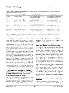
Table 1. Impact of heparan sulfate proteoglycan (HSPG) and β-amyloid (Aβ) interactions on amyloid deposition and tissue
properties in Alzheimer’s disease (AD)
HSPG Expression in AD Interactions with Aβ Effects on tissues
Perlecan Elevated levels in AD 56 Inhibits amyloid degradation in vitro and Perlecan domain V inhibits α2β1 and
57
promotes amyloid deposition α5β1 integrin-mediated Aβ toxicity 58-61
Agrin Agrin is abundant in Aβ plaques and Binding of agrin to Aβ increases and Elevated endothelial agrin Aβ deposition
elevated in CSF of AD patients stabilizes fibril formation regulates BBB permeability
GPC 1-6 GPC 1-6 are downregulated in the Aβ treatment of neurons and astrocytes Alterations in the distribution of GPC4
cerebellum but upregulated in limbic amplifies GPC1 transcription and sulphation; lead to cognitive and social disorders 63
areas in AD. 62 GPC4 aids Aβ internalization and toxicity
SDC1 – 4 Hippocampus SDC3 and SDC4 are Neuronal SDC3 plays a critical role in Aβ1 SDC3 contributes to long-term
elevated in AD, corresponding with – 42 endocytosis 64 potentiation in the hippocampus 65
disease severity
Collagen XVIII Upregulated expression in the Endostatin forms cytotoxic amyloid Dysfunctional ECM in AD is linked
(COL18A1) entorhinal cortex and hippocampus fibrils in vitro; Coll XVIII forms vascular to COL18A1 mutations, which lead
66
in severe AD 40 amyloid deposits and senile plaques in to Knobloch syndrome and brain
AD. 67 abnormalities
Abbreviations: BBB: Blood–brain barrier; CSF: Cerebrospinal fluid; ECM: Extracellular matrix; GPC: Glypican; LRP-1: Low-density lipoprotein
receptor-related protein-1; SDC: Syndecan.
abnormally folded α-syn, all of which detrimentally perlecan have been shown to associate with Aβ and α-syn
impact neuronal viability and function. Parkinson’s aggregates, thereby promoting their propagation and
19
disease (PD), the second most common neurodegenerative persistence in AD and PD. 30
disorder after AD, is characterized by a loss of functional
brain dopaminergic neurons in the brain, leading to 5.1. Inter-α-trypsin inhibitor (ITI)/bikunin and
impaired motor control. Aggregated forms of α-syn are related Kunitz domain proteins in brain tissues
abundant in pathological PD tissues and are involved in ITI is localized in the nuclei of neurons and astrocytes
its pathogenesis. Numerous studies have suggested that across all age groups, with cytoplasmic expression
27
HSPG interactions with tau contribute to various stages of becoming more prominent in adulthood. Members of
31
the development of AD, including seeding, aggregation, the ITI family are components of the innate immune
plaque formation, neurofibrillary tangle-mediated toxicity, system within the CNS and exhibit neuroprotective
31
and delayed clearance of amyloid deposits. The oligomeric properties. In neonatal rats, ITI levels decrease following
28
32
Aβ fragment activates hyperphosphorylation of the tau hypoxic-ischemic brain injury. Bikunin, the light chain
protein. In a healthy brain, tau maintains axonal stability, of ITI, contains two KPI domains that inhibit pancreatic
ensuring efficient transmission of action potentials during trypsin, chymotrypsin, leukocyte elastase, and cathepsin
neurotransduction. However, hyperphosphorylation of tau G, kallikrein, and plasmin. Bikunin has been identified in
disrupts axonal transmission, leading to cytotoxic signaling. the hippocampus and cerebral cortex and has also been
Recent research has proposed that HSPG-tau assemblies detected in human brain tumors. Other related Kunitz
33
contribute to the formation of tunneling nanotubes in domain proteins include tissue factor pathway inhibitors
brain tissues, facilitating the intercellular transport of α, β, and δ, as well as the multidomain transmembrane
tau and Aβ peptides. These nanotubes play a role in the serine protease inhibitors HAI-1 and HAI-2 (hepatocyte
seeding of amyloid deposits and the pathogenesis of AD. 29
growth factor [HGF] activator inhibitors 1 and 2).
5. Central nervous system (CNS) proteins These multidomain proteins are neuroprotective and
with Kunitz protease inhibitor (KPI) modules membrane-bound and play roles in maintaining the
integrity of the neural basement membrane and brain
in the delayed clearance of pathological microvasculature, in addition to exhibiting anticoagulant
protein aggregates in AD and PD activity.
CNS proteoglycans containing core proteins KPI modules 5.2. Tissue factor protease inhibitors (TFPI) 1 and 2
possess tissue-protective properties. However, when these in the brain
proteoglycans are associated with pathological protein
aggregates, they may impede the clearance of deposits TFPIα is a protein weighing approximately 43 kDa and
observed in AD and PD. HSPGs such as agrin and contains three tandem KPI domains. The TFPIβ isoform
Volume 3 Issue 3 (2024) 4 doi: 10.36922/an.3812