
Page 67 - AN-3-3
P. 67
Advanced Neurology mTOR inhibition in epilepsy
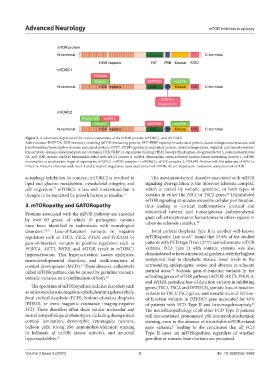
Figure 2. A schematic depiction of the main components of the mTOR protein, mTORC1, and mTORC2
Abbreviations: DEPTOR: DEP-domain-containing mTOR-interacting protein; FAT: FKBP-rapamycin-associated protein, ataxia-telangiectasia mutated, and
transformation/transcription-domain-associated protein; FATC: FKBP-rapamycin-associated protein, ataxia-telangiectasia mutated, and transformation/
transcription-domain-associated protein C-terminal; FRB: FKBP 12-rapamycin-binding; HEAT repeats: Huntington, elongation factor 3, protein phosphatase
2A, and TOR repeats; mLST8: Mammalian lethal with SEC13 protein 8; mSIN1: Mammalian stress-activated protein kinase interacting protein 1; mTOR:
mammalian or mechanistic target of rapamycin; mTORC1: mTOR complex 1; mTORC2: mTOR complex 2; PRAS40: Proline-rich Akt substrate of 40 kDa;
Protor ½: Proteins observed with rictor 1 and 2; Raptor: Regulatory-associated protein of mTOR; Rictor: Rapamycin-insensitive companion of mTOR.
autophagy inhibition. In contrast, mTORC2 is involved in The most prototypical disorder associated with mTOR
lipid and glucose metabolism, cytoskeletal integrity, and signaling dysregulation is the tuberous sclerosis complex,
cell migration. mTORC2 is less well understood but is which is caused by somatic, germline, or both types of
17
20
thought to be regulated by growth factors or insulin. 17 variants in either the TSC1 or TSC2 genes. Disinhibited
mTOR signaling stimulates excessive cellular proliferation,
3. mTORopathy and GATORopathy thus leading to cortical malformations (cortical and
Proteins associated with the mTOR pathway are encoded subcortical tubers) and tumorigenesis (subependymal
by over 60 genes, of which 16 pathogenic variants giant cell astrocytomas or hamartomas in other organs) in
20
have been identified in individuals with neurological tuberous sclerosis complex.
disorders. 16,18 Loss-of-function variants in negative Focal cortical dysplasia Type II is another well-known
21
regulators such as TSC1, TSC2, PTEN, and STRADA or mTORopathy. Lim et al. found that 15.6% of the studied
gain-of-function variants in positive regulators such as patients with FCD type II (n=12/77) carried somatic mTOR
PI3KCA, AKT3, RHEB, and MTOR, result in mTORC1 variants. FCD Type II with somatic variants was also
hyperactivation. This hyperactivation causes epilepsies, demonstrated to have a mutational gradient, with the highest
neurodevelopmental disorders, and malformations of mutational load in dysplastic tissues, lower levels in the
cortical development (MCD). These diseases, collectively surrounding epileptogenic zones, and absence in adjacent
16
22
called mTORopathies, can be caused by germline variants, normal areas. Somatic gain-of-function variants in the
somatic variants, or a combination of both. 16 activating genes of mTOR pathway (MTOR, AKT3, PIK3CA,
and RHEB), germline loss-of-function variants in inhibiting
The spectrum of mTORopathies includes disorders such genes (TSC1, TSC2, and DEPDC5), somatic loss-of-function
as tuberous sclerosis, megalencephaly, hemimegalencephaly, variants in TSC1/TSC2 genes, and somatic second-hit loss-
focal cortical dysplasia (FCD), bottom-of-sulcus dysplasia of-function variants in DEPDC5 gene accounted for 63%
(BOSD), or even magnetic resonance imaging-negative of patients with FCD Type II and hemimegalencephaly.
23
FCD. These disorders often share similar molecular and The neurohistopathology of all other FCD Type II patients
neurohistopathological phenotypes, including disorganized still demonstrated pronounced pS6 immunohistochemical
cortical lamination, dysmorphic cytomegalic neurons, staining, even in the absence of identifiable mTOR-related
balloon cells, strong pS6 immunohistochemical staining gene variants, leading to the conclusion that all FCD
23
(a hallmark of mTOR kinase activity), and neuronal Type II cases are mTORopathies, regardless of whether
hyperexcitability. 19 germline or somatic brain variants are presented.
Volume 3 Issue 3 (2024) 3 doi: 10.36922/an.3568