
Page 156 - AN-3-4
P. 156
Advanced Neurology Non-invasive electroencephalography in rats
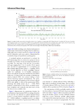
A
B
Figure 4. Examples of spike-wave activity induced by xylazine (i.p., 2 mg/kg) in a symptomatic WAG/Rij rat (female, 16 months old). The time of injection
was designated as time zero. (A) Continuous SWDs were recorded from symmetrical frontal (left and right) and occipital right cortical locations using
epidural ECoG. (B) Continuous SWDs recorded using the EEG technique in the same rat subject 40 days before ECoG.
Abbreviations: ECoG: Electrocorticography; SWD: Spike-wave discharges; WAG/Rij: Wistar Albino Glaxo from Rijswijk.
(Figure 4B). Both recordings were obtained subsequent to
xylazine injections at a dose of 2 mg/kg. Note the consistency
of the spike-wave activity’s waveform in Figure 4A and 4B
and the distinctive spike-wave pattern, characterized by
remarkably sharp and repetitively occurring spikes.
A statistical analysis was performed to evaluate the
SWD-promoting effect of xylazine in a group of 16 rats
(9 females and 7 males). All rats were implanted with ECoG
electrodes. The mean age of the rats was 14.4 months,
with ages ranging from 13 to 16 months. ECoG signals
were recorded for approximately 24 h at baseline and
after xylazine administration (dose 2 – 8 mg/kg). SWDs
were visually identified in ECoG recordings obtained
under two conditions: (1) At baseline: spontaneous
SWDs were selected during the 4-h interval (from 0:00 to Figure 5. Pearson correlation between the total duration of spontaneous
04:00 a.m.). (2) Immediately after xylazine administration: SWDs occurring at baseline (4 h) and the total duration of xylazine-
xylazine-induced SWDs were selected during a 6-min induced SWDs (6 min).
interval following xylazine administration. The spike- Abbreviation: SWD: Spike-wave discharges.
wave morphology and frequency of the xylazine-induced
SWDs were similar to those of the spontaneous SWDs. total duration of xylazine-induced SWDs, indicating a
Following this initial period, the 8 – 10-Hz SWDs high level of consistency between them. The occurrence
gradually transitioned to 6 Hz SWDs, occasional spike- of xylazine-induced SWDs was found to be minimal in
wave complexes, and eventually into slow-wave activity subjects with no spontaneous SWDs at baseline (Figure 5).
that characterizes a sedative state. The total duration of SWDs was highly variable among
Figure 5 demonstrates the correlations between the the 16 rat subjects. Four rats (25% from the group, marked
total duration of spontaneous SWDs occurring at baseline with brown dots in Figure 5) did not experience any SWD
(4 h) and the total duration of xylazine-induced SWDs during the baseline period, and they exhibited minimal
(6-min). A strong Pearson correlation coefficient of 0.72 spike-wave activity following xylazine administration
(P = 0.0016) indicates a significant positive correlation (<25 s out of a 300-s observation period, or <8%). Therefore,
between the total duration of spontaneous SWDs and the these subjects were considered asymptomatic. The other
Volume 3 Issue 4 (2024) 6 doi: 10.36922/an.4464