
Page 117 - AN-4-3
P. 117
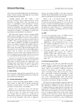
Advanced Neurology                                                     Subcortical fibers in band heterotopia


            with a more severely affected phenotype than heterozygous   behavior  and  reduced  mobility  of  her  right  extremities.
            women, wherein neurons expressing the non-mutated   Her EEG showed bifrontal spike-wave discharges. The scan
            gene might migrate appropriately.                  had to be performed under general anesthesia.

              Clinically, patients with SBH exhibit a wide       Patient 2 was a 22-year-old woman who started
            spectrum of features. Typical symptoms include mental   experiencing tonic–clonic convulsions at the age of
            retardation  and  high  seizure  rates,  depending  on  the   12 years and presented with episodes of “loss of look and
            degree of manifestation of the heterotopia.  Invasive   absentia.” Her EEG revealed frequent interictal spike-wave
                                                 4,5
            electroencephalogram (EEG), single photon emission   discharges. Under lamotrigine and valproic acid treatment,
            computed tomography, and experimental data suggest   her seizure rate could be reduced to 2 – 5 episodes/month.
            that the heterotopia and overlying cortex contribute to the   She had no neurologic or neurologic deficit and worked
            development of epilepsy.  Accordingly, no specific EEG   successfully as a psychologist.
                                6-9
            patterns have been described in the literature. 10
                                                               2.2. MRI
              Early  and recent magnetic  resonance  imaging  (MRI)
            studies using tractography have shown a rather “isolated”   All scans were performed using a 3T Philips Achieva
            band caused by interruptions of central or more peripheral   scanner and included the following sequences:
            white matter tracts at the inner respective outer border of   •   T1-weighted: 3D turbo field echo MPRAGE, repetition
            the SBH. 11-13  Thus, epileptic discharges from the overlying   time/echo time (TR/TE) = 6.73/3.11 ms, number of
                                                                                                            3
            cortex might be difficult to propagate to generalization.   sagittal slices = 180, reconstructed voxel size = 1 mm .
            However, according to histology, this “isolation” does not   •   DTI: Number of gradient directions = 32, b = 0 and
            appear to be real. “All subtypes of inhibitory GABAergic   800  s/mm²,  measured voxel  size =  2 × 2  × 2  mm,
            interneurons intermingled with pyramidal neurons were   number of slices covering the whole head = 60, SENSE
            observed in the white matter. This network of differentiated   factor = 2.
            neurons with its dendritic and axonal ramifications   •   Functional MRI  (patient  2 only):  Echo  planar
            suggests the presence of functional neuronal circuitry, and   imaging–gradient echo sequence: TR = 2000 ms,
            there is strong observational evidence that the two cortices   number  of slices  with 3-mm  thickness =  34, gap =
            were anatomically and functionally interconnected.” 14, p.1838  0.75 mm, field of view = 192 mm, voxel size = 2.38 ×
                                                                  2.38 × 2 mm, and number of slices covering the whole
              Owing to this ostensible discrepancy between imaging   head = 34.
            and post-mortem findings, we examined the subcortical
            white matter (SCWM) between the malformation and   2.3. Post-processing of data
            overlying cortex and between the central white matter   Post-processing of DTI data was performed using the
            (CentWM) and longer tracts as well as explored the   program  “MRtrix3”  (https://www.mrtrix.org),  supported
            organization of the malformation using diffusion tensor   by “FSL” (FMRIB Software Library, https://fsl.fmrib.ox.ac.
            imaging (DTI)–based tractography.                  uk/fsl). MRtrix uses multishell multitissue constrained

            2. Case series                                     spherical deconvolution to improve fiber tracking in
                                                               voxels containing crossing fibers and partial volume
            The retrospective study had been approved by the local   effect, anatomically constrained tractography to reject
            ethics committee of CEDIMAT, and informed consent was   streamlines ending in biologically implausible tissues
            obtained from each participant.                    such as the cerebrospinal fluid (CSF), and spherical
                                                               deconvolution informed filtering of tractograms, which
            2.1. Controls and patients 1 and 2                 corrects the overestimation of longer streamlines.
            Overall, 2 patients with drug-resistant epilepsy, in whom   DTI data from images arranged in stacks were denoised
            MRI revealed an SBH, and 10 healthy age-matched    and corrected for ringing artifacts and EPI distortion, and
            volunteers (control group), who had undergone MRI   fiber distribution orientation was then estimated. The
            previously for other reasons, were enrolled.       T1-weighted images were segmented into tissue types such

              Patient 1 was a 24-year-old woman who had been   as gray matter, CentWM, and CSF and coregistered to the
            suffering from tonic and rarely from clonic seizures since   DTI. For general tractography, streamlines were calculated
            the age of 13  years. Her seizure rate initially reached   from the coregistered gray matter–white matter border
            up to 50/day, which was reduced by a combination of   templates for the whole brain. Tracts within the SCWM
            carbamazepine, valproic acid, and lacosamide. Cognitively,   were edited from these streamlines by excluding all tracts
            she  had  mental  retardation  with  spells  of  aggressive   that entered into the CentWM. Vice versa, CentWM tracts


            Volume 4 Issue 3 (2025)                        111                               doi: 10.36922/an.4823