
Page 120 - AN-4-3
P. 120
Advanced Neurology Subcortical fibers in band heterotopia
A B Table 1. Number of subcortical and central white matter
tracts and DTI parameters of these regions as well as those
from the heterotopias
Patient Patient 2 Control Control
1 (Mean) (Standard
deviation)
No. of SCWM tracts 502225* 476515* 3251372.2* 1329843.11
No. of CentWM tracts 471004 585255 734084.4 180669.19
FA SCWM 0.194 0.228 0.204 0.0259
FA CentWM 0.285 0.37 0.322 0.0232
2
MD SCWM (mm /s) 1.145 0.959 1.063 0.1666
2
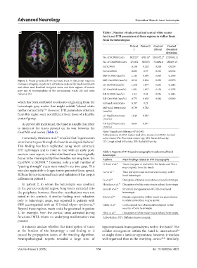
Figure 5. Tracts generated from activated areas in functional magnetic MD CentWM (mm /s) 0.916 0.804 0.829 0.0970
resonance imaging. In patient 2, activations induced by hand movements LD SCWM (mm /s) 1.354 1.177 1.235 0.1414
2
and vision were localized in typical areas, and their regions of interest 2
gave rise to tractographies of the corticospinal tracts (A) and optic LD CentWM (mm /s) 1.201 1.137 1.124 0.1235
radiation (B). RD SCWM (mm /s) 1.04 0.85 0.936 0.1013
2
RD CentWM (mm /s) 0.773 0.638 0.684 0.0869
2
which has been attributed to seizures originating from the FA band heterotopia 0.187 0.23
heterotopic gray matter that might exhibit “altered white MD band heterotopia 0.739 0.788
matter connectivity.” However, DTI parameters obtained (mm /s)
16
2
from this region were not different from those of a healthy LD band heterotopia 1.046 0.983
control group. (mm /s)
2
As previously mentioned, the band is usually described RD band heterotopia 0.665 0.691
2
to interrupt the tracts present on its way between the (mm /s)
CentWM and cortex (Table 2). Note: *Significant difference (P=0.030).
Abbreviations: SCWM: Subcortical white matter; CentWM: Central
Conversely, Erickson et al. revealed that “eigenvectors white matter; FA: Fractional anisotropy; MD: Mean diffusivity;
17
were found to pass through the band in an aligned fashion.” LD: Longitudinal diffusivity; RD: Radial diffusivity.
This finding has been replicated using more advanced
DTI techniques and is more in line with an early post- Table 2. Reports of DTI‑based tractography in subcortical band
mortem case report, in which the band was histologically heterotopia
found to be interrupted by fiber bundles running from the Authors Main findings related to DTI tractography
CentWM to SCWM. However, only a small number of Erikson et al. 17 Tracts may pass or end within the bands, and fibers
18
“passing-through” tracts were noted in our two cases. This may originate from the bands
was also applicable to longer tracts generated from special Lee et al. 11 Tract disruption and decreased anisotropy within
ROIs as the corticospinal tracts and radiation of the corpus band heterotopia
callosum in patient 1. Lu et al. 12 Disruption of frontal connections in band heterotopia
In patient 2, in whom the heterotopia was confined Hoischen et al. 19 Disruption of white matter tracts in band heterotopia
to the parieto-occipital region, long tracts extended into Iannetti et al. 16 Structural disorganization of U-fibers in band
the periphery; however, these fine ramifications were not heterotopia
noted in the controls. A similar finding, tract reduction Kini et al. 24 Neurite organization within bands was more similar
only in heterotopic areas, was reported in patients with to white matter than to gray matter
SBH accompanied with an X-linked Alport syndrome. Chiba et al. 5 Corticospinal tract abnormalities depend on the
19
Beyond these regions, tracts could be generated in patient severity of band heterotopia
2, for example, from the cortical areas activated during Zhou et al. 13 Disruption of white matter tracts in band heterotopia
functional MRI, where no underlying malformation was Abbreviation: DTI: Diffusion tensor imaging.
present.
It remains unclear whether this interruption of tracts hypomyelinated brain parenchyma within the band. The
4
at the borders of the heterotopy a real finding or is cellular arrangement within the band is unstructured
14
caused by propagation issues of the tracking algorithm. or might show a laminar appearance; however, it was less
Neuropathological reports revealed a large zone of well-organized than in the overlying cortex. 20,21 Similarly,
Volume 4 Issue 3 (2025) 114 doi: 10.36922/an.4823