
Page 119 - AN-4-3
P. 119
Advanced Neurology Subcortical fibers in band heterotopia
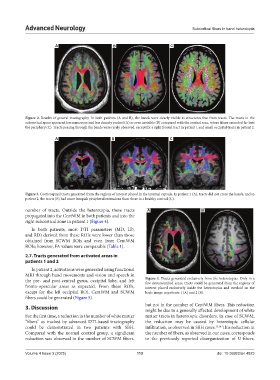
A B C
Figure 2. Results of general tractography. In both patients (A and B), the bands were clearly visible as structures free from tracts. The tracts in the
subcortical space appeared less numerous and less densely packed (A) or even invisible (B) compared with the control scan, where fibers extended far into
the periphery (C). Tracts passing through the bands were rarely observed, except for a right frontal tract in patient 1 and small occipital tracts in patient 2.
A B C
Figure 3. Corticospinal tracts generated from the regions of interest placed in the internal capsule. In patient 1 (A), tracts did not cross the bands, and in
patient 2, the tracts (B) had more lumpish peripheral extensions than those in a healthy control (C).
number of tracts. Outside the heterotopia, these tracts A B
propagated into the CentWM in both patients and into the
right subcortical zone in patient 1 (Figure 4).
In both patients, most DTI parameters (MD, LD,
and RD) derived from these ROIs were lower than those
obtained from SCWM ROIs and even from CentWM
ROIs; however, FA values were comparable (Table 1).
2.7. Tracts generated from activated areas in
patients 1 and 2
In patient 2, activations were generated using functional
MRI through hand movements and vision and speech in
the pre- and post-central gyrus, occipital lobe, and left Figure 4. Tracts generated exclusively from the heterotopias. Only in a
few circumscribed areas, tracts could be generated from the regions of
fronto-opercular areas as expected. From these ROIs, interest placed exclusively inside the heterotopias and overlaid on the
except for the left occipital ROI, CentWM and SCWM basic image in patients 1 (A) and 2 (B).
fibers could be generated (Figure 5).
3. Discussion but not in the number of CentWM fibers. This reduction
might be due to a generally affected development of white
For the first time, a reduction in the number of white matter matter tracts in heterotopic disorders. In case of SCWM,
“fibers” as tracked by advanced DTI-based tractography the reduction may be caused by heterotopic cellular
could be demonstrated in two patients with SBH. infiltration, as observed in SBH cases. 13,14 This reduction in
Compared with the normal control group, a significant the number of fibers, as observed in our cases, corresponds
reduction was observed in the number of SCWM fibers, to the previously reported disorganization of U-fibers,
Volume 4 Issue 3 (2025) 113 doi: 10.36922/an.4823