
Page 98 - AN-4-3
P. 98
Advanced Neurology TG100-115 suppresses glioblastoma cell functions
240 µM), revealing significant damage and the presence of following treatment with either DMSO (0.1%) or TG100-
visualized vacuoles when compared to the control group 115 (150 µM), and the wound closure rate was analyzed. At
(Figure 2A, n = 4). This suggests that TG100-115 treatment 4, 8, 16, and 24 h, the control group exhibited wound closure
resulted in morphological changes in the U251 cells. rates of 14.8 ± 1.4%, 37.4 ± 1.4%, 48.3 ± 1.8%, and 66.1 ±
2.6%, respectively, all of which were notably faster than
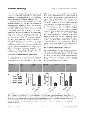
To assess apoptosis, we conducted western blot analysis the TG100-115-treated group at the corresponding time
of cleaved caspase-3, Bcl-2, and Bax. The representative points: 8.6 ± 1.2%, 20.2 ± 2.3%, 28.3 ± 3.4%, and 41.5 ± 2.2%
images are shown in Figure 2B, the raw blot images are (p<0.01, p<0.001, n ≥ 6). This result suggests a significant
provided in Figures S5–8. Cleaved caspase-3, an active and time-dependent reduction in the migratory capability
form of the apoptosis-related cysteine peptidase, showed of U251 cells treated with TG100-115 when compared to
25
a significantly higher ratio in TG100-115 (150 µM)- the control group. In Figure 4, we further investigated the
treated cells (34.5 ± 2.6%) compared to the control group migration of U251 cells with various doses of TG100-115 (60,
(6.4 ± 0.7%, p<0.01, n = 4), as demonstrated in Figure 2C. 120, 180 µM) for 24 h, using DMSO (0.1%) as the control.
26
Bcl-2 and Bax, pivotal regulatory proteins in apoptosis, The wound closure rate in the control group was 66.1 ± 2.6%,
were also analyzed. Bcl-2, an anti-apoptotic protein, while in the treatment groups, it was significantly reduced
exhibited a significantly lower ratio in TG100-115 (150 to 48.8 ± 3.7%, 43.2 ± 3.7%, and 37.4 ± 1.4%, respectively
µM)-treated cells (14.2 ± 5.3%) compared to the control (p<0.01, p<0.001, n ≥ 6). This result indicates a significant
group (48.5 ± 3.8%, p<0.05, n = 4), as shown in Figure 2D. reduction in the migration of U251 cells treated with TG100-
Conversely, Figure 2E illustrates a significant increase in Bax 115 compared to the control across different doses.
level (97.4 ± 13.3%) after treatment with TG100-115 (150
µM) for 24 h, compared to the control group (45.7 ± 9.3%, 3.4. TG100-115 inhibited U251 cell invasion
p<0.05, n = 4). These findings collectively suggest that The Matrigel transwell assay was utilized to assess U251
TG100-115 induces apoptosis in U251 cells. cell invasion. As depicted in Figure 5, U251 cell invasion
was significantly reduced after 24-h exposure to TG100-
3.3. TG100-115 suppressed U251 cell migration
115 at 150 µM. The relative invasiveness decreased from
To evaluate the impact of TG100-115 on the migratory 100.0 ± 8.8% to 53.8 ± 3.8% with TG100-115 treatment
behavior of U251 cells, a wound healing assay was performed. (p<0.01, n = 6). This result indicates that, in vitro, TG100-
In Figure 3, cell images were captured at 0, 4, 8, 16, and 24 h 115 diminishes the invasion of U251 cells.
A
B C D E
Figure 2. TG100-115 induced apoptosis in U251 cells. (A) Representative images of U251 cells with or without TG100-115 (30 – 240 µM) treatment
for 24 h. Scale bar: 50 µm, magnification ×20. U251 cells treated with TG100-115 significantly showed cell damage and the presence of vacuoles
when compared to the control group. (B) Representative images of western blotting results. (C) TG100-115 significantly increased the ratio of cleaved
caspase-3/GAPDH. (D) TG100-115 significantly reduced the ratio of Bcl-2/GAPDH. (E) TG100-115 significantly increased the ratio of Bax/GAPDH.
Note: Statistical significance determined at *p<0.05 and **p<0.01 using Student’s t-test, n = 4.
Abbreviations: Bax: B-cell lymphoma 2-associated X protein; Bcl-2: B-cell lymphoma 2; DMSO: Dimethyl sulfoxide; GAPDH: Glyceraldehyde-3-
phosphate dehydrogenase.
Volume 4 Issue 3 (2025) 92 doi: 10.36922/AN025110023