
Page 96 - ARNM-2-4
P. 96
Advances in Radiotherapy
& Nuclear Medicine Malignant peripheral nerve sheath tumor: A case report
A B exhibit any other adverse effects, such as infection,
thrombocytopenia, nausea, vomiting, or hair loss.
However, after three chemotherapy cycles, the patient
experienced bilateral lower limb weakness, left-side
dominance, left cranial nerve VI paralysis, and blurred
vision in the left eye. The patient underwent contrast-
enhanced cranial MRI, which revealed an 18 × 31-mm
lesion ventral to the pons, with mixed signals and strong
C D and heterogeneous enhancements after contrast injection,
and thickening of the adjacent meninges (Figure 6).
The physician explained that the disease was progressing
despite chemotherapy; however, the patient and her family
refused to continue treatment and chose palliative care and
symptomatic management in medical facilities.
3. Discussion
1
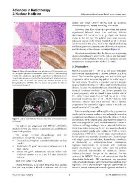
Figure 5. Histopathological features on microscope. (A). Low-power view MPNSTs account for 5 – 10% of all soft-tissue sarcomas
of a malignant peripheral nerve sheath tumor (MPNST) demonstrating and occur in approximately 1/100.000 individuals in both
variable hypocellular and hypercellular areas. (B and C). Moderate-power sexes. This disease has a poor prognosis and exhibits rapid
3
view showing elongated, spindle-like shaped cells with a hyperchromatic progression, often metastasizing primarily to the lungs in
nucleus, with cell necrosis and mitosis are common features in the
histopathology in MPNSTs. (D). The tumor was positive for S100 in the early stages. To achieve a negative resection margin,
immunohistochemistry. surgical treatment is the only radical treatment for localized
disease. In cases of distant metastasis, chemotherapy is an
essential treatment modality. The disease generally has
a poor prognosis, with an overall 5-year survival rate of
15 – 66%, 5-year event-free survival rate of 24 – 53%,
1
1
and local recurrence rate of 20 – 85.7%. Patients with
1
metastatic disease have poor survival, with a median
progression-free survival of approximately 4 months and
overall survival of 13 months. 2
Patients usually present with a rapidly enlarging mass that
may be painful or cause local neurological symptoms such as
weakness or paresthesias. In most cases, the mass is >5 cm at
Figure 6. Lesions anterior to the pons are metastatic nerve sheath tumors presentation. In the present case, the patient was diagnosed
4
(red arrow)
with a late-stage MPNST, with multiple metastatic lesions in
The patient was diagnosed with MPNST cT4N0M1, the bones and lungs leading to severe pain and debilitation.
multifocal bone and bilateral lung metastases, and bilateral Pubic bone lesion biopsy results and immunohistochemical
lower limb paralysis. staining revealed spindle cells positive for S100, a protein
The patient received chemotherapy with the characteristic of MPNSTs. The only viable treatment option
doxorubicin–ifosfamide–mesna regimen: for the patient was systemic chemotherapy. Chemotherapy
• Doxorubicin 30 mg/m ; intravenous infusion on days with single agents (dacarbazine, doxorubicin, epirubicin,
2
1 and 2. or ifosfamide) or anthracycline-based combination
• Ifosfamide 3.75 g/m ; intravenous infusion over 4 h, regimens (doxorubicin or epirubicin with ifosfamide
2
days 1 and 2. and/or dacarbazine) has been widely used for patients
5
• Mesna 750 g/m ; intravenous infusion before each with advanced, unresectable, or metastatic disease.
2
ifosfamide session and at 4 and 8 h after ifosfamide Anthracycline-based therapy is the standard first-line
6
infusion. treatment for MPNST. Kroep et al. demonstrated that the
Each cycle lasted for 21 days. doxorubicin–ifosfamide (AI) combination was associated
with a lower risk of relapse and better response rate than other
During treatment, the patient developed mild anemia regimens. Some chemotherapy regimens appropriate for
7
that did not require a blood transfusion and did not MPNSTs, mainly anthracycline-based regimens, are listed in
Volume 2 Issue 4 (2024) 3 doi: 10.36922/arnm.3462