
Page 95 - ARNM-2-4
P. 95
Advances in Radiotherapy
& Nuclear Medicine Malignant peripheral nerve sheath tumor: A case report
defecation or urination disorders. No special signs were lesion exposed uniform spin hyalinization stroma. The
noted on clinical examination, except for paralysis in both tumor stained positive for S100 in immunohistochemistry
lower extremities, with muscle strength of 4/5. (Figure 5). These features indicated a malignant epithelioid
Her blood cell counts and blood biochemistry test results peripheral nerve sheath tumor.
were within the normal range. Magnetic resonance imaging The patient also underwent esophagogastroduodenoscopy
(MRI) of the lumbar and sacral spine revealed multiple and colorectal endoscopy, which revealed no abnormal lesions
hyperintense lesions in T2-weighted imaging (T2WI) and suggestive of malignancy.
fluid-attenuated inversion recovery, rim enhancement in
the vertebral body, and vertebral pedicle on both sides of L4. A B
The lesions, including a large 22 × 12-mm focus, exhibited
substantial spinal membrane and soft-tissue infiltration,
causing nerve compression at the L4 level. The area of
damage to the left L2 pedicle was comparable, measuring
14 × 17 mm. The D11–D12 vertebral body had foci of
hyperintense lesions on T2WI and strong gadolinium
enhancement. The invasive D12 lesion damaged the bone
cortex and grew into the spinal canal (Figure 1).
Thoracic computed tomography revealed several solid Figure 2. Chest computed tomography scan showing solid nodules
nodules with well-circuited rounded lesions of varying scattered in the lung fields on both sides. (A) Solid nodule (red arrow) in left
sizes and heterogeneous gadolinium enhancements upper lobe lung. (B) Solid nodule in the right lower lobe lung (blue arrow)
scattered on both sides of the lungs (Figure 2). The bone and some nodules with the same characteristic in the left lung.
lesions in right rib V, rib arches IV and VI, and bilateral
sternoclavicular joint caused bone destruction and A B
adjacent soft-tissue invasion (Figure 3A-C). Abdominal
computed tomography revealed two nodules with poor
contrast enhancement in the right and left liver lobes. No
malignant cells were found in the biopsy of the right lobe
nodule (Figure 3D).
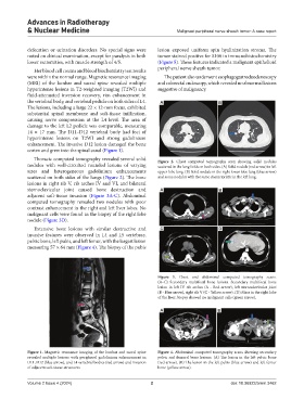
Extensive bone lesions with similar destructive and C D
invasive features were observed in L4 and L5 vertebrae,
pelvic bone, left pubis, and left femur, with the largest lesion
measuring 57 × 64 mm (Figure 4). The biopsy of the pubis
Figure 3. Chest and abdominal computed tomography scans.
(A–C) Secondary multifocal bone lesions. Secondary multifocal bone
lesion in left IV rib arches (A - Red arrow), left sternoclavicular joint
(B - Blue arrow), right rib V (C - Yellow arrow). (D) Mass in the right lobe
of the liver; biopsy showed no malignant cells (green arrow).
A B
Figure 1. Magnetic resonance imaging of the lumbar and sacral spine Figure 4. Abdominal computed tomography scans showing secondary
revealed multiple lesions with peripheral gadolinium enhancement in pelvic and femoral bone lesions. (A) The lesion in the left pelvic bone
D11, D12 (blue arrow), and L4 vertebral bodies (red arrow) and invasion (red arrow). (B) The lesion in the left pubis (blue arrow) and left femur
of adjacent soft-tissue structures bone (yellow arrow).
Volume 2 Issue 4 (2024) 2 doi: 10.36922/arnm.3462