


Page 46 - ARNM-3-3
P. 46
Advances in Radiotherapy
            & Nuclear Medicine                                                PI3K and Akt in androgen-independent PCa


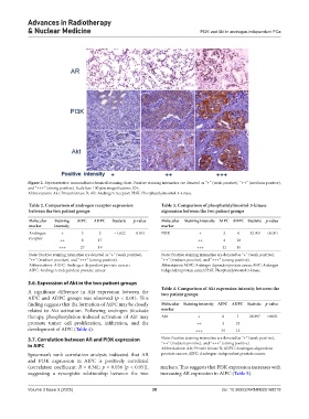
            Figure 2. Representative immunohistochemical staining chart. Positive staining intensities are denoted as “+” (weak positive), “++” (medium positive),
            and “+++” (strong positive). Scale bar: 100 μm; magnification: 20×.
            Abbreviations: Akt: Protein kinase B; AR: Androgen receptor; PI3K: Phosphatidylinositol-3-kinase.

            Table 2. Comparison of androgen receptor expression   Table 3. Comparison of phosphatidylinositol‑3‑kinase
            between the two patient groups                     expression between the two patient groups
            Molecular   Staining   AIPC  ADPC  Statistic  p‑value  Molecular   Staining intensity AIPC ADPC Statistic p‑value
            marker    intensity                                marker
            Androgen     +      3      2     −1.622  0.105     PI3K           +        2     0   32.901  <0.001
            receptor    ++      8      17                                    ++        4    28
                        +++     27     19                                    +++       32   10
            Note: Positive staining intensities are denoted as “+” (weak positive),   Note: Positive staining intensities are denoted as “+” (weak positive),
            “++” (medium positive), and “+++” (strong positive).  “++” (medium positive), and “+++” (strong positive).
            Abbreviations: ADPC: Androgen-dependent prostate cancer;    Abbreviations: ADPC: Androgen-dependent prostate cancer; AIPC: Androgen-
            AIPC: Androgen-independent prostate cancer.        independent prostate cancer; PI3K: Phosphatidylinositol-3-kinase.

            3.6. Expression of Akt in the two patient groups
                                                               Table 4. Comparison of Akt expression intensity between the
            A significant difference in Akt expression between the   two patient groups
            AIPC and ADPC groups was observed (p  < 0.01). This
            finding suggests that the formation of AIPC may be closely   Molecular  Staining intensity AIPC  ADPC  Statistic  p‑value
            related to Akt activation. Following androgen blockade   marker
            therapy, phosphorylation-induced activation of Akt may   Akt     +         0    1    28.897  <0.001
            promote tumor cell proliferation, infiltration, and the          ++        3    24
            development of AIPC (Table 4).                                  +++       35    13

            3.7. Correlation between AR and PI3K expression    Note: Positive staining intensities are denoted as “+” (weak positive),
            in AIPC                                            “++” (medium positive), and “+++” (strong positive).
                                                               Abbreviations: Akt: Protein kinase B; ADPC: Androgen-dependent
            Spearman’s  rank  correlation analysis  indicated that  AR   prostate cancer; AIPC: Androgen-independent prostate cancer.
            and PI3K expression in AIPC is positively correlated
            (correlation coefficient: R = 0.341; p = 0.036 [p < 0.05]),   markers. This suggests that PI3K expression increases with
            suggesting a synergistic relationship between the two   increasing AR expression in AIPC (Table 5).


            Volume 3 Issue 3 (2025)                         38                        doi: 10.36922/ARNM025160018

