


Page 45 - ARNM-3-3
P. 45
Advances in Radiotherapy
            & Nuclear Medicine                                                PI3K and Akt in androgen-independent PCa


                                 A                    B                     C


                                 D                    E                     F


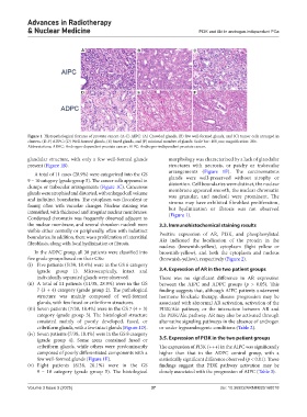
            Figure 1. Histopathological features of prostate cancer. (A-C) AIPC: (A) Crowded glands, (B) few well-formed glands, and (C) tumor cells arranged in
            clusters. (D-F) ADPC: (D) Well-formed glands, (E) fused glands, and (F) minimal number of glands. Scale bar: 100 μm; magnification: 20×.
            Abbreviations: ADPC: Androgen-dependent prostate cancer; AIPC: Androgen-independent prostate cancer.

            glandular structure, with only a few well-formed glands   morphology was characterized by a lack of glandular
            present (Figure 1B).                                  structures with necrosis, or patchy or trabecular
                                                                  arrangements (Figure  1F).  The carcinomatous
              A total of 11 cases (28.9%) were categorized into the GS
            9 – 10 category (grade group 5). The cancer cells appeared in   glands were well-preserved without atrophy or
                                                                  distortion. Cell boundaries were distinct, the nuclear
            clumps or trabecular arrangements (Figure 1C). Cancerous   membrane appeared smooth, the nuclear chromatin
            glands were atrophied and distorted, with enlarged cell volume
            and indistinct boundaries. The cytoplasm was flocculent or   was granular, and nucleoli were prominent. The
            foamy,  often  with  vacuolar  changes.  Nuclear  staining  was   stroma may have exhibited fibroblast proliferation,
                                                                  but hyalinization or fibrosis was not observed
            intensified, with thickened and irregular nuclear membranes.   (Figure 1).
            Condensed chromatin was frequently observed adjacent to
            the nuclear membrane, and several shrunken nucleoli were   3.3. Immunohistochemical staining results
            visible either centrally or peripherally, often with indistinct
            boundaries. In addition, there was a proliferation of interstitial   Positive expression of AR, PI3K, and phosphorylated
            fibroblasts, along with focal hyalinization or fibrosis.  Akt indicated the localization of the protein in the
                                                               nucleus (brownish-yellow), cytoplasm (light yellow or
              In the ADPC group, all 38 patients were classified into   brownish-yellow), and both the cytoplasm and nucleus
            five grade groups based on their GSs:              (brownish-yellow), respectively (Figure 2).
            (i)  Five patients (5/38, 18.4%) were in the GS 6 category
               (grade group  1). Microscopically, intact and   3.4. Expression of AR in the two patient groups
               individually separated glands were observed.    There was no significant difference in AR expression
            (ii)  A total of 11 patients (11/38, 28.9%) were in the GS   between the AIPC and ADPC groups (p  > 0.05). This
               7 (3 + 4) category (grade group 2). The pathological   finding suggests that, although AIPC patients underwent
               structure  was  mainly  composed  of  well-formed   hormone blockade therapy, disease progression may be
               glands, with few fused or cribriform structures.  associated with abnormal AR activation, activation of the
            (iii) Seven patients (7/38, 18.4%) were in the GS 7 (4 + 3)   PI3K/Akt pathway, or the interaction between AR and
               category (grade  group  3). The  histological structure   the PI3K/Akt pathway. AR may also be activated through
               consisted mainly of poorly developed, fused, or   alternative signaling pathways in the absence of androgen
               cribriform glands, with a few intact glands (Figure 1D).  or under hypoandrogenic conditions (Table 2).
            (iv)  Seven patients (7/38, 18.4%) were in the GS 8 category
               (grade group  4). Some areas contained fused or   3.5. Expression of PI3K in the two patient groups
               cribriform glands, while others were predominantly   The expression of PI3K (+++) in the AIPC was significantly
               composed of poorly differentiated components with a   higher than that in the ADPC control group, with a
               few well-formed glands (Figure 1E).             statistically significant difference observed (p < 0.01). These
            (v)  Eight patients (8/38, 20.1%) were in the GS   findings suggest that PI3K pathway activation may be
               9  –  10 category (grade group  5). The histological   closely associated with the progression of AIPC (Table 3).


            Volume 3 Issue 3 (2025)                         37                        doi: 10.36922/ARNM025160018

