
Page 24 - BH-1-1
P. 24
Brain & Heart Cerebral venous thrombosis mimicking brain tumors
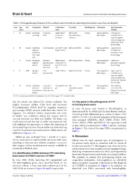
Table 1. Clinicopathological features of five cerebral venous thrombosis mimicking brain tumor cases from our hospital
Case Age Sex Symptoms History Laboratory Location Misdiagnosis Treatment Intraoperative
(year) tests finding
1 23 Female Headache, Caesarean High platelet Left frontal Glioma Anticoagulation Left superior
vomiting birth count and lobe and corpus after biopsy cerebral vein to
D-dimer callosum superior sagittal
sinus drainage was
obstructed, focal
hemorrhage
2 48 Female Headache, Oral ibuprofen Low APTT and Left parietal lobe Glioma Anticoagulation Large branch
epilepsy, high D-dimer after biopsy of Labbé vein
numbness infarction, focal
over right hemorrhage
arm
3 13 Female Headache, Excessive High platelet Left temporal lobe Glioma Anticoagulation Labbé vein
epileptic menstruation count and TT; after biopsy infarction, regional
seizure treated by low APTT and hemorrhage
Marvelon Fib
4 31 Male Epileptic Oral High D-dimer Right frontal lobe Glioma Anticoagulation Right superior
seizure paracetamol after biopsy cerebral vein to
superior sagittal
sinus drainage was
obstructed, regional
hemorrhage
5 35 Female Headache, Long-term Hemoglobin<60 Left Glioma Anticoagulation The draining
dizziness moderate to g/L temporal-occipital after biopsy vein of the left
incontinence severe anemia lobe temporal cortex was
(twice) blue-black
APTT: Activated partial thromboplastin time; Fib: Fibrinogen; TT: Thrombin time
that the edema was induced by venous occlusion. The 3.3. Key genes in the pathogenesis of CVT
slightly increased choline (Cho) level and decreased mimicking brain tumor
N-acetylaspartate (NAA) level by magnetic resonance In total, 38 genes were related to thrombophilia, as
spectroscopy (MRS) analysis could have also misled the demonstrated by the MalaCards human disease database.
diagnosis. An infarcted inferior anastomotic vein (vein According to the differential gene criteria Q-value ≤ 0.05
of Labbé) was confirmed during the surgery and its and FC ≥ 2 or FC ≤ 0.5, the following nine differential genes
vascular structure was dark and swollen. The lesion was were screened: SERPINE1, SELP, THBD, ITGB3, TFPI,
totally resected and the vein of Labbé was preserved well F13A1, PROS1, PPBP, and PROCR. The expression levels
with sufficient decompression to relieve the symptoms of of nine DEGs are presented in Table 2 and in a heatmap
intracranial hypertension. Hematoxylin and eosin staining in Figure 5. The roles of the nine DEGs are presented in
revealed thrombosed vein and excessive inflammatory cell Table 3.
infiltration (Figures 1-3).
Follow-up was continued from 1 month to 5 years. 4. Discussion
All the study subjects had favorable outcomes with intact Glioma was the most common type of misdiagnosis in
neurological function and without symptom recurrence the present study, which is consistent with the results of
after surgery. Follow-up information was not available in the previous studies [7,15] . Misdiagnosis was observed in the
previously published cases. present study due to non-specific primary imaging findings,
including enhancement and mass effect, accompanied by
3.2. Identification of DEGs between CVT mimicking intracranial hypertension and similar neurological deficits.
brain tumor (CVTMBT) and non-CVTMBT
The majority of patients had predisposing factors and
In total, 1066 DGEs, including 813 upregulated and coagulation dysfunction. Anticoagulation can effectively
253 downregulated genes, were identified based on the improve patients’ prognosis. A flowchart for the diagnosis
selection criteria. A heat map and a volcano plot of the of CVTMBT is presented in Figure 6. In addition, we
1066 DGEs are shown in Figure 4A and B, respectively. identified nine DEGs associated with thrombophilia,
Volume 1 Issue 1 (2023) 4 https://doi.org/10.36922/bh.v1i1.188