
Page 25 - BH-1-1
P. 25
Brain & Heart Cerebral venous thrombosis mimicking brain tumors
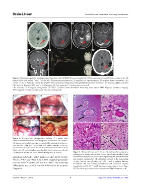
A B C D
E F G
Figure 1. Typical pre-operative imaging images of a patient with CVTMBT from our hospital. (A) CT scan showing low-density lesion located in the left
temporal lobe (red arrow). (B and C) Axial MRI demonstrating isointense on T1-weighted and hyperintense on T2-weighted lesion, respectively (red
arrow). (D and E) Axial and coronal T1-weighted MRI showing no enhancement after gadolinium injection (red arrow). (F) Showing slightly increased
Cho level and decreased NAA level by MRS analysis. (G) Post-operative CT showing lesion removed.
Cho: Choline; CT: Computed tomography; CVTMBT: Cerebral venous thrombosis mimicking brain tumor; MRI: Magnetic resonance imaging;
MRS: Magnetic resonance spectroscopy; NAA: N-acetylaspartate
A B A B
C D
C D
Figure 2. Representative intraoperative findings of a patient with
cerebral venous thrombosis mimicking brain tumor from our hospital.
(A) Intraoperative photo showing obvious Labbé vein infarct (red arrow
indicates the Labbe vein), with dark and swollen vascular structure.
(B) Red arrow indicates the occlusion involved in the main branch of the
Labbé vein. (C) Lesion totally removed and the Labbé vein preserved well.
(D) Normal structure and blood flow of drainage vein as control. Figure 3. Hematoxylin and eosin (H and E) staining of post-operative
pathology of a patient with cerebral venous thrombosis mimicking brain
including SERPINE1, SELP, THBD, ITGB3, TFPI, F13A1, tumor from our hospital. (A) H and E staining showing two thrombosed vein
PROS1, PPBP, and PROCR, by mRNA sequencing between and excessive inflammatory cell infiltration scattered in the surrounding
patients with CVTMBT and non-CVTMBT. The screening of vein (original magnification ×10). (B) H and E staining showing
thrombosed vein and excessive inflammatory cell infiltration (original
of key genes may provide valuable information for accurate magnification ×40). (C) H and E staining showing regional hemorrhage.
diagnosis. (D) Vacuolar degeneration of nerve tissue enclosed by inflammatory cell.
Volume 1 Issue 1 (2023) 5 https://doi.org/10.36922/bh.v1i1.188