
Page 55 - BH-1-2
P. 55
Brain & Heart UVC-related infective endocarditis and septic emboli
Daily blood cultures consistently yielded positive the neonate underwent 2 weeks of triple antibiotic therapy
results for MSSA, despite the administration of an (nafcillin, rifampin, and gentamicin) and 4 additional
appropriate antibiotic regimen. Repeat echocardiogram weeks of dual antibiotic therapy with nafcillin and rifampin
showed increased size of thrombus or vegetation for endocarditis involving prosthetic material caused
(9.7 mm × 4.7 mm) with no valvular involvement. by staphylococci. He was discharged home on DOL 56.
Considering the challenge of achieving effective source His discharge included a regimen encompassing aspirin,
control and the concerns regarding potential septic emboli LMWH, and levetiracetam. On the 3-month follow-up
reaching both the brain and lungs in the settings of elevated assessment, the neonate had remained clinically stable
pulmonary pressures, the decision was made to proceed and been on full feeds with no evidence of intracardiac
with surgical intervention. thrombus. Moreover, his cardiac and neurological function
On DOL 10, the neonate underwent an atrial septectomy, remained commendable.
accompanied by the debridement of LA and the right-sided 3. Discussion
pulmonary veins and an atrial reconstruction using a Gore-
Tex patch. This surgical intervention was followed by the The umbilical vein is a common site for neonatal central
removal of the UVC. Postoperatively, the neonate required venous access to administer medications and parenteral
vasoactive support involving epinephrine and vasopressin nutrition (PN), as well as to obtain blood for laboratory
[5]
for 4 days. Throughout this period, the administration of studies . However, the use of UVCs is not without
antibiotics was continued, culminating in the achievement complications. Among these, serious complications include
of the first negative blood culture outcome on DOL 13. encompass bloodstream infections, thromboembolism,
The neonate was successfully extubated to a high-flow air embolism, arrhythmia, hydrothorax, hemorrhage,
[5]
nasal cannula on DOL 14. Commencing on DOL 15, malposition, and migration . This case report highlighted
the neonate’s medical regimen encompassed aspirin, the successful surgical management of IE in the setting
introduced for patch prophylaxis. A new PICC line was of a malpositioned UVC that resulted in systemic septic
inserted on DOL 18, replacing the old one. Subsequently, thrombosis.
on DOL 20, the anticoagulation regimen was transitioned The predominant UVC-related complications are
from UFH to therapeutic low-molecular-weight heparin bloodstream infections, with reported rates ranging from
(LMWH), with an anti-factor Xa assay (anti-Xa) goal 3% to 36%, depending on the applied diagnostic criteria .
[5]
range of 0.35 – 0.7 U/milliliter. Since the neonates with UVCs are not routinely screened
The ophthalmologic examination yielded no ocular for thrombus formation, current rates and clinical
signs secondary to infective endocarditis. Ultrasound significance of UVC-related thrombosis remain obscure.
evaluations of the abdomen, head, and lower extremities The incidences of UVC thrombosis vary widely, spanning
ruled out the presence of possible abscesses. A subsequent from 3% to 33% in historical series encompassing surviving
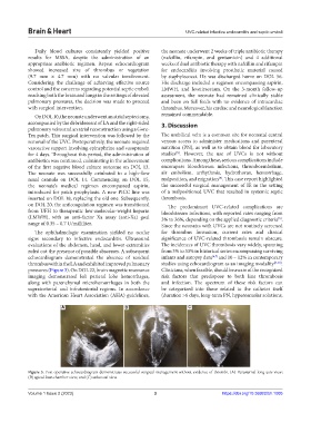
echocardiogram demonstrated the absence of residual infants and autopsy data [6,7] and 10 – 12% in contemporary
thrombus within the LA and exhibited improved pulmonary studies using echocardiogram as an imaging modality [8-10] .
pressures (Figure 3). On DOL 22, brain magnetic resonance Clinicians, when feasible, should be aware of the recognized
imaging demonstrated left parietal lobe hemorrhages, risk factors that predispose to both line thrombosis
along with parenchymal microhemorrhages in both the and infection. The spectrum of these risk factors can
supratentorial and infratentorial regions. In accordance be categorized into those related to the catheter itself
with the American Heart Association (AHA) guidelines, (duration >6 days, long-term PN, hyperosmolar solutions,
A B C
Figure 3. Post-operative echocardiogram demonstrates successful surgical management without evidence of thrombi. (A) Parasternal long-axis view;
(B) apical four-chamber view; and (C) subcostal view.
Volume 1 Issue 2 (2023) 3 https://doi.org/10.36922/bh.1005