
Page 54 - BH-1-2
P. 54
Brain & Heart UVC-related infective endocarditis and septic emboli
infection can activate platelets, fostering conditions
favorable for thrombus formation [1-4] . Most of these
complications typically resolve with the removal of the
catheter, supplemented by anticoagulation and antibiotic
therapies . Surgical intervention, on the other hand,
[3]
remains a rarity . In this case report, we present the
[4]
management approach employed for a term neonate with
an intracardiac thrombus, infective endocarditis, and
subsequent septic emboli related to a malpositioned UVC.
2. Case presentation
A male neonate born at full term with a birthweight of
2950 g was transferred to our hospital at 6 days of life (DOL)
for the management of UVC-related IE. His birth was
uncomplicated, marked by vaginal delivery, with APGAR
scores of 8 and 9. However, by DOL 2, he developed Figure 1. Initial chest X-ray demonstrates the umbilical venous catheter
hypoglycemia and respiratory distress, prompting his tip terminating above the cavoatrial junction.
transfer to the neonatal intensive care unit at an outlying
facility. A UVC was inserted on DOL 2 and the initial sepsis A B
workup, along with blood cultures, was not concerning for
infection. However, on DOL 4, due to feeding intolerance,
a repeat sepsis workup was performed, revealing
leukopenia, thrombocytopenia, elevated inflammatory
markers, and coagulopathy. In response, blood cultures
were sent, and a treatment regimen involving ampicillin
and gentamicin was initiated. Lumbar puncture was C D
deferred due to coagulopathy. A head ultrasound (HUS)
yielded results within normal limits. Chest X-ray revealed
the presence of a UVC situated at a high position inside
the right atrium (Figure 1), which was not repositioned.
On DOL 5, blood culture results indicated the presence
of gram-positive cocci, prompting the addition of
vancomycin to the treatment regimen. In the setting of the
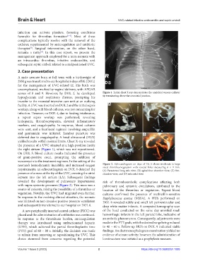
neonate’s hemodynamic instability and increased oxygen Figure 2. Echocardiogram on days of life 6 shows moderate-to-large
requirements, an echocardiogram on DOL 6 detected the sized thrombus/vegetation with several lobes measuring 6 × 9 mm.
(A) Parasternal long axis view; (B) apical four-chamber view; (C) five-
presence of a mass at the tip of the UVC, crossing the atrial chamber view; and (D) subcostal view.
septum into the left atrium (LA). Subsequent findings
revealed the development of pulmonary hypertension risk of thromboembolic complications affecting both
with supra-systemic pressures (Figure 2). This mass was a pulmonary and systemic circulations, attributed to the
source of concern, raising the possibility of a thrombus or location of the thrombus or vegetation. Repeat blood
vegetation. Notably, the UVC had migrated even further. cultures confirmed the presence of methicillin-sensitive
In response to the evolving clinical situation, the neonate Staphylococcus aureus (MSSA). A HUS performed on
was initiated on non-invasive positive pressure ventilation DOL 8 revealed subtle and small left periventricular and
and subsequently transferred to our hospital on DOL 6. deep white matter infarcts. A computed tomography scan
A new peripherally inserted central catheter (PICC) was of the head conducted on the same day unveiled small
placed and the administration of antibiotics was continued. hemorrhagic infarcts in the left parietal lobe, indicative of
In response to the thrombosis burden, anticoagulation an embolic phenomenon. Consequently, adjustments were
therapy was introduced using unfractionated heparin made to the PTT goals, with the desired range being reduced
(UFH), which achieved the partial thromboplastin time to 40 – 60 s. Follow-up HUS on DOL 9 indicated stable
(PTT) goal of 60 – 80 s. Initially, the decision was made findings. An electroencephalogram examination yielded no
to refrain from removing or repositioning the UVC. This evidence of seizure activity; however, the administration of
choice stemmed from concerns regarding the potential levetiracetam was initiated as a prophylaxis measure.
Volume 1 Issue 2 (2023) 2 https://doi.org/10.36922/bh.1005