
Page 75 - BH-2-1
P. 75
Brain & Heart Lutembacher’s percutaneous treatment
2. Case presentation pressure of 50 mmHg. The MV commissures were free
of significant calcification, and no significant subvalvular
A 30-year-old male presented with complaints of progressive thickening was noted. These findings, along with adequate
dyspnea on exertion, easy fatiguability, and occasional ASD rim margins, suggested an opportunity for definitive
non-exertional palpitations over the past 4 years, with a percutaneous management. After obtaining informed
worsening of symptoms in the past 6 months. There were consent as per institutional guidelines, the patient
no reported orthopnea or paroxysmal nocturnal dyspnea underwent BMV, followed by percutaneous ASD device
episodes. On cardiovascular examination, notable findings closure in the same sitting.
included a loud S1, the presence of a wide fixed-split S2, a
loud P2 component, a mid-diastolic murmur at the apex The patient received an oral dose of aspirin (325 mg)
lacking pre-systolic accentuation, and a Grade 2 ejection and clopidogrel (300 mg) 1 day before the procedure, in
7
systolic murmur in the second left intercostal space. accordance with the guidelines. Intravenous heparin was
administered at an initial dose of 5000 IU after obtaining
The 12-lead electrocardiogram indicated sinus rhythm,
bi-atrial enlargement, right ventricular hypertrophy, and peripheral access, with a repeat dose given after 1 h to
maintain the activated clotting time over 200 s (as the
a right bundle branch block pattern. In addition, the procedure lasted for an unexpectedly long duration).
chest X-ray revealed a straightened left heart border, a Right femoral venous access was secured, and a Mullin’s
double atrial shadow sign, and borderline cardiomegaly dilator (8F sheath) was used to insert a 035” hydrophilic
with pulmonary venous congestion and dilated proximal Terumo wire through the ASD into the LA. At this stage,
pulmonary artery segments. the mean LA pressure was 6 mmHg, while the mean right
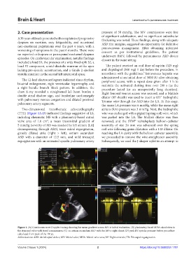
Two-dimensional transthoracic echocardiography atrium (RA) pressure was 3 mmHg. Next, the hydrophilic
(TTE) (Figure 1A-D) indicated findings suggestive of LS, wire was exchanged with a pigtail (spring coil) wire, which
including rheumatic MS with a planimetry-based mitral was parked into the LA. The Mullin’s dilator was then
valve area of 1.0 cm , a mean transmitral gradient of removed, and the SYM valvuloplasty balloon-catheter
2
5 mmHg (severity of MS was masked by left atrium [LA] assembly of size 26 mm was advanced over the spring
decompressing through ASD), trace mitral regurgitation, coil wire following groin dilatation with a 14F dilator. On
grossly dilated atria (right > left), ostium secundum reaching the LA cavity with the balloon catheter assembly,
ASD with a diameter of 12.5 mm, and mild tricuspid we proceeded to remove the wire-straightener assembly.
regurgitation with an estimated systolic pulmonary artery Subsequently, we used the J-shaper stylet in an attempt to
A B
C D
Figure 1. (A) Continuous wave Doppler tracing showing the mean gradient across MV at initial evaluation; (B) planimetry-based MVA calculation in
the stenosed valve with fused commissures; (C) an ostium secundum ASD with the left to right shunt; (D) peak RV systolic pressure before procedure
calculated from peak of the TR jet.
Abbreviations: ASD: Atrial septal defect; MV: Mitral valve; MVA: Mitral valve area; RV: Right ventricle; TR: Tricuspid regurgitation.
Volume 2 Issue 1 (2024) 2 https://doi.org/10.36922/bh.1701