
Page 80 - BH-2-1
P. 80
Brain & Heart Pacing strategy for heart block
A C
B
Figure 1. Baseline electrocardiogram indicating complete right bundle
branch block.
into the right ventricle. A SelectSecure lead (Model 3830;
Medtronic, Inc., Minnesota, USA) was then screwed
into the RV septum. The pacing lead was maneuvered to
the tagged area, in which unipolar electrode tip pacing
demonstrated QRS complex with a “W pattern” in ECG
lead V and RS pattern in the inferior lead. As the lead was
1
advanced, the W shape in lead V changed to a QR pattern
1
with a QRS duration of 158 ms (Figure 2). Notably, no
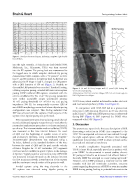
discernible LBB potential was recorded. Threshold testing, Figure 2. (A and B) X-ray images and (C) paced QRS during left
utilizing unipolar pacing, revealed left ventricular septum ventricular septal pacing.
pacing (LVSP) without LBB capture, consistent with the Abbreviations: LAO: Left anterior oblique; LVS: Left ventricular septum;
criteria established by Wu et al. The pacing parameters RAO: Right anterior oblique.
2
were within acceptable ranges (pacemaker sensing:
10 mV; pacing threshold: 0.4 mV/0.4 ms, and pacing 2.0 V/0.4 ms, which resulted in favorable cardiac electrical
impedance: 580 Ω). An unexpectedly narrower QRS of and mechanical synchrony (Table 1 and Figure 3).
non-RBBB morphology was noticed when a bipolar pacing In comparison with LVSP, BSP led to a pronounced
configuration was adopted. This finding indicated that reduction in QRS duration. However, no R’ was observed
BSP was achieved with the capture of both aspects of the in ECG lead V (QRS with RBBB pattern was not observed)
1
septum when bipolar pacing was performed. during BSP (Figure 4). BSP improved the IVMD when
ECG measurements were obtained using a multichannel compared with LVSP (Figure 5).
recorder. Echocardiography was performed 1 week after the
pacemaker implantation procedure to evaluate mechanical 3. Discussion
synchrony. The interventricular mechanical delay (IVMD) The present case report is the first case description of BSP,
was measured as the time interval between the onset showcasing a reduction in IVMD time compared to the
of QRS and the beginning of systolic waves of aortic LVSP. This unexpected achievement was realized through
and pulmonary ejections, using conventional Doppler. the right septal capture with an LVS lead. Our findings
3
Intraventricular dyssynchrony was evaluated using a underscore the potential of BSP to attain optimal cardiac
Yu index, defined as the standard deviation of the time electrical and mechanical synchrony.
between the onset of QRS and the peak systolic velocity A notable complication frequently associated with
of tissue Doppler for 12 left ventricular (LV) segments TAVR is heart block, especially prevalent in patients with
(six basal and six middle) in apical triplane-mode (using a prior RBBB. As documented in several studies, chronic
4
4-Vprobe). Septal-posterior wall motion delay (SPWMD) RV pacing is linked to an increased risk of heart failure.
3
4
was measured through M-mode as the distance between Recently, LBBP has emerged as a new pacing approach.
5
the first maximum systolic inward motion of the septum However, while LBBP produces a relatively narrow QRS
and the maximum inward motion of the posterior wall. complex with fast left ventricular activation, concerns
Anodal capture using the ring electrode in the persist regarding the presence of RBBB or even incomplete
RV septum was observed using bipolar pacing at RBBB during pacing, leading to potential interventricular
Volume 2 Issue 1 (2024) 2 https://doi.org/10.36922/bh.1670