
Page 82 - BH-2-1
P. 82
Brain & Heart Pacing strategy for heart block
A B C D
Figure 4. Electrocardiogram measurements. (A) Left ventricular activation time. (B) QRS duration. (C) Delayed right ventricular activation time. (D) QRS
morphology in lead V .
1
Abbreviations: BSP: Bilateral septal pacing; LVSP: Left ventricular septal pacing.
A B comparable levels of electrical and mechanical activation
in the left ventricle. Second, our findings, based on
9,10
ECG characteristics and echocardiography analyses, were
limited by the absence of information on activation patterns.
Further investigation should incorporate endocardial
activation mapping to provide a more comprehensive
understanding of cardiac activation during pacing. Third,
the inability to achieve anode capture in certain cases,
C D attributed to a small length of 3830 lead penetration into
the septum or a high capture threshold, highlights the need
for the development of new electrodes to overcome these
challenges.
As previously stated, our study assessed synchrony
during BSP and LVS pacing in a patient with a heart block.
While we believe our findings have broader relevance,
future studies involving a larger cohort will play a pivotal
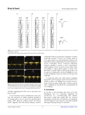
Figure 5. Interventricular mechanical delay during bilateral septal pacing role in validating the reproducibility and generalizability
(BSP) and left ventricular septal pacing (LVSP). The images show the time
to onset of left ventricular ejection during LVSP (A) and BSP (C) and the of our findings.
time to onset of right ventricular ejection during LVSP (B) and BSP (D).
4. Conclusion
SPWMD, suggesting that LVSP induces interventricular To the best of our knowledge, this report is the first
dyssynchrony. investigation into the mechanism underlying the
Several limitations warrant consideration in our study. phenomenon of a narrowed-paced QRS duration
First, our assessment of cardiac synchrony focused on and reduced IVMD time during BSP. The observed
LVS pacing, and we did not explore the synchronization maintenance of both inter- and intraventricular synchrony
during LBBP. However, existing animal and human with BSP suggests it could represent a more physiologically
studies suggested that both pacing strategies maintain advantageous pacing strategy for heart block.
Volume 2 Issue 1 (2024) 4 https://doi.org/10.36922/bh.1670