
Page 81 - BH-2-1
P. 81
Brain & Heart Pacing strategy for heart block
Table 1. Analyses of cardiac electrical and mechanical synchrony
Electrocardiogram Echocardiography
QRSd (ms) V 1 a LVAT (ms) dRVAT (ms) IVD (ms) IVMD (ms) Ts‑SD (ms) b PSD c SPWMD (ms)
BSP 114 QS 78 ‑ ‑ 12 61.5±3.7 85 103
LVSP 158 QR 82 126 42 43 96±2.0 101 132
Notes: QRS morphology in lead V ; Ts-SD: Yu index (standard deviation of the time to longitudinal peak strain of 17 segments); PSD: Peak strain
a
c
b
1
dispersio7n.
Abbreviations: BSP: Bilateral septal pacing; dRVAT: delayed right ventricular activation time (the interval from the pacing to the peak R’ wave upstroke
in ECG lead V ); IVD: Interventricular conduction delay (dRVAT-LVAT); IVMD: Interventricular mechanical delay; LVAT: Left ventricular activation
1
time (the interval from the pacing to the peak R wave upstroke); LVSP: Left ventricular septal pacing; QRSd: QRS duration; SPWMD: Septal-posterior
wall motion delay.
A B
C
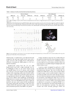
Figure 3. Electrocardiogram and schematic of activation pattern during bilateral septal pacing. (A) Paced QRS. (B) The red arrow indicates the ring of the
pacing lead. (C) Schematic of the activation pattern.
1,6
dyssynchrony. A study by Lin et al.6 demonstrates that Cardiac mechanical synchrony was notably enhanced
pacing both left and right bundle branch areas with a during BSP, as demonstrated by echocardiography analyses.
single ventricular lead can further shorten QRS duration, Cardiac synchrony is important for maintaining cardiac
effectively eliminating the RBBB pattern on ECG in a structure and function, and cardiac resynchronization has
majority of patients. been identified as a valuable strategy for mitigating the risk
Simultaneously pacing both the left and right of heart failure events. 7
bundle branch areas with a single ventricular lead can A previous study investigated the application of BSP
be challenging at times. In our patient, bipolar pacing in combination with coronary venous pacing for cardiac
with anodal capture of the RV septum could attenuate resynchronization therapy. The study revealed that the
8
interventricular synchrony. Unipolar pacing at high output combination of BSP with coronary venous pacing resulted
yielded a QRS morphology without any transitions before in superior acute electrical synchronization compared to
loss of capture, indicating left ventricular septal capture by conventional cardiac resynchronization therapy. However,
the ring electrode without LBB capture. Bipolar pacing at the study did not incorporate mechanical synchronization
2.0 V/0.4 ms yielded a QRS morphology, eliminating the as a parameter. Echocardiography offers an accurate and
RBBB pattern and characterized by a relatively narrow QRS convenient method for evaluating cardiac contraction and
complex, which was attributable to RV septum capture by hemodynamics in real time without exposing the patient
anodal. In our case, anodal capture by the ring electrode to radiation. In our patient, we employed IVMD as an
was possible only in the myocardium, as the initial ECG index for interventricular synchrony and SPWMD as an
exhibited RBBB. BSP appeared to achieve more favorable index for intraventricular synchrony. Our observation
cardiac electrical synchrony than LBBP in our patient. revealed that LVSP distinctly prolonged IVMD and
Volume 2 Issue 1 (2024) 3 https://doi.org/10.36922/bh.1670