
Page 85 - BH-2-1
P. 85
Brain & Heart Prosthesis-sparing aortic root replacement
consultation from us complained of dull chest pain. inserted in the false lumen and secured with BioGlue
Echocardiography showed a marked dilatation of the (CryoLife Europa Inc., Surrey, UK) in the previous operation
sinus of Valsalva, and a hyperechoic line suggesting a was solely exposed (Figure 2A). Remnant BioGlue was
polyester fabric sheet inserted in the dissected aortic root seen around the polyester fabric. An approximately 10 mm
in the previous operation. The prosthetic valve function intimal tear, which was not resulted from the previous
was normal with mean transvalvular pressure gradient operation, appeared in the non-coronary sinus. The
of 7 mmHg, and there was no paravalvular leakage proximal aortic suture line of the previous operation was
(Figure 1A). Computed tomography revealed a huge intact. The 25-mm REGENT mechanical valve (Abbott,
pseudoaneurysm of the aortic root reaching beneath CA, USA) was left in place, and a total of 16 2-0 bladed
the sternum (Figure 1B and C). His blood pressure was mattress sutures were circumferentially placed in the
138/80 mmHg, and heart rate 77 beats/min on admission. sewing ring of the mechanical valve. A 28-mm Valsalva
The patient underwent an urgent operation. Before redo graft (Terumo Aortic, FL, USA) was cut at the middle of
sternotomy, cardiopulmonary bypass was started with its skirt portion, through which these sutures were passed
right femoral vein drainage with a 25 Fr venous reuptake and tied, and was seated to completely cover the cuff of the
cannula and right femoral artery perfusion using a 9-mm mechanical valve (Figure 2C and D). The right coronary
Dacron graft attached to the femoral artery. Then, the apex button was created and sutured to a 11-mm Dacron graft
of the heart was exposed through the left 5 intercostal to approximate the intima and the adventitia because the
th
space, and a left ventricular apical vent was inserted, aortic dissection involved the right coronary sinus. The
followed by systemic cooling. Redo sternotomy was left coronary sinus was free from aortic dissection, but
performed during hypothermic ventricular fibrillatory the left coronary artery was difficult to mobilize due to
arrest at systemic temperature of 22°C. After the redo severe adhesion. Therefore, an 11-mm graft was attached
sternotomy was completed, the mediastinal adhesions to the orifice of the left coronary artery using the inclusion
were dissected. The pseudoaneurysm was immediately technique. These grafts were attached to the Valsalva graft
entered, leading to a massive bleeding. Hypothermic using the Piehler technique, through which the coronary
circulatory arrest was induced, and the adhesion around arteries were reconstructed with graft interposition. Lastly,
the ascending aortic graft was removed. Following this, the Valsalva graft was anastomosed to the previous 26-mm
the graft was securely cross-clamped and transected. Then, ascending graft to complete the procedure. The patient was
systemic perfusion was resumed, and rewarming was discharged home without complications.
started, followed by selective antegrade cardioplegic arrest.
The right main pulmonary artery was lacerated due to the 3. Discussion
adhesion of the ascending graft, and it was repaired with a In patients with a history of AVR, aortic root dissection or
bovine pericardial patch. root dilation may sometimes occur. However, this aortic
The whole adventitia of the non-coronary sinus pathology is not caused by the implanted aortic prosthetic
disappeared, and the polyester fabric which had been valve. Dilatation or dissection of the aortic root constitutes
A B C
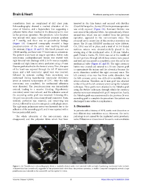
Figure 1. (A) Transthoracic echocardiography shows a markedly dilated aortic root (asterisk) with normally functioning mechanical valve (arrow).
A hyperechoic line (arrowhead), suggestive of the polyester fabric sheet from the previous operation, is visible. Computed tomography reveals a large
pseudoaneurysm (asterisk) in the coronal (B) and the sagittal (C) views.
Volume 2 Issue 1 (2024) 2 https://doi.org/10.36922/bh.2256