
Page 24 - BH-2-4
P. 24
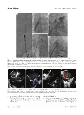
Brain & Heart                                                      Step by step 3D ICE guided LAA occlusion


                         A                     B                      D


                                               C

                                                                      E


            Figure 1. Fluoroscopy views. (A) J Wire in the RA provides a “road map” for facilitating ICE advancement through the iliac veins and inferior vena cava
            under ICE guidance. (B) Interatrial septostomy may be needed for difficult ICE catheter crossing across the IAS. (C) Flossing the puncture site with the
            Watchman FLX sheath first and proper alignment of the ICE catheter with the transeptal wire are important steps to facilitate easier passage of the ICE
            catheter across the IAS. (D) Position of ICE catheter on fluoroscopy for left superior pulmonary vein view of the LAA. (E) Position of ICE catheter on
            fluoroscopy for supramitral view of the LAA.
            Abbreviations: RA: Right atrium; ICE: Intracardiac echocardiography; IAS: Interatrial septum; LAA: Left atrial appendage.

            A                        B                        C                         D


            Figure 2. ICE views from RA. (A) Home view, (B) Left atrial appendage, (C) Left-sided pulmonary veins, (D) Right-sided pulmonary veins.
            Abbreviations:  RA:  Right  atrium;  TV:  Tricuspid  valve;  RV:  Right  ventricle;  MV:  Mitral  valve;  LAA:  Left  atrial  appendage;  IAS:  Interatrial  septum;
            LPV: Left-sided pulmonary veins; RPV: Right-sided pulmonary veins; ICE: Intracardiac echocardiography.

            c.  If  necessary,  balloon septostomy with  an 8–10-mm   2.1.6. ICE views in LA
               Mustang balloon may be employed to slightly     a.  Once the probe reaches the LA, three views can be
               expand the puncture site during the septal crossing   used. However, due to 3D MPR capability, the entire
               (Figure 1B).                                       procedure can also be performed in a single view.


            Volume 2 Issue 4 (2024)                         5                                doi: 10.36922/bh.4018