
Page 26 - BH-2-4
P. 26
Brain & Heart Step by step 3D ICE guided LAA occlusion
position, compression, and leakage. It is crucial to b. To check for pericardial effusion, use the RA home
conduct a careful 360° evaluation to assess for the view at a lower magnification. Multiplane imaging is
peri-device leak (PDL), which can be readily achieved preferred. If there is any concern, RV ICE views can be
using the 4D ICE probe. used to evaluate the pericardial space beyond the LV
and RV.
2.1.8. ICE views from RA
a. Once the LAAO device is released and the final 2.1.9. Access site care
assessment is completed, withdraw the probe into the a. The ICE probe is then removed along with the sheath
RA to evaluate the IAS at the TSP site for iatrogenic b. Hemostasis is achieved with vascular closure device
ASD evaluation (size and shunt) like Perclose, figure of 8 stitch or manual compression.
A B 2.2. Procedural outcomes
Both groups achieved a technical success rate of 100%,
which is defined as successful device deployment without
embolization and a PDL of <3 mm. No devices were
recaptured in Group 2 (all procedures were accomplished
using one device and in one attempt), whereas device
recapture was necessary for one case in Group 1; however,
it did not alter the final size of the Watchman FLX
deployed. In Group 1, the average duration of the entire
C D procedure was 102.0 ± 37.7 min, whereas in Group 2, it
was 89.0 ± 11.4 min (Table 3). The average duration from
ICE insertion to septal crossing was 16 min, and the
average duration from septal crossing to LAAO device
deployment was 30 min in Group 2. The fluoroscopy
duration improved in Group 2 (24.9 min) compared with
that in Group 1 (28.4 min). One patient in each group
underwent pre-planned patent foramen ovale (PFO)
closure during the LAAO procedure to diminish the
overall risk of embolic stroke. The total procedure time
Figure 5. ICE views in LA. (A) Left atrial “home view.” (B) Left reflects the additional time it took to perform PFO closure
superior pulmonary vein view, which is analogous to the 0° TEE view.
(C) Supramitral view, which is similar to the 135° TEE view. (D) Aortic as well. None of the patients in either group required iASD
valve view, which is comparable to the 45° TEE view. closure.
Abbreviations: LAA: Left atrial appendage; AV: Aortic valve;
ICE: Intracardiac echocardiography; LA: Left atrium, TEE: Transesophageal The most frequent device size implemented under
echocardiography. 3D ICE guidance was 27 mm; no resizing was required.
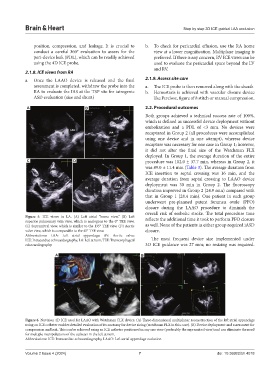
A B
Figure 6. Nuvision 4D ICE used for LAAO with Watchman FLX device. (A) Three-dimensional multiplanar reconstruction of the left atrial appendage
using an ICE catheter enables detailed evaluation of its anatomy for device sizing (watchman FLX in this case). (B) Device deployment and assessment for
compression and leak. This can be achieved using an ICE catheter positioned in any one view (preferably the supramitral view) and can eliminate the need
for multiple manipulations of the catheter in the left atrium.
Abbreviations: ICE: Intracardiac echocardiography, LAAO: Left atrial appendage occlusion.
Volume 2 Issue 4 (2024) 7 doi: 10.36922/bh.4018