
Page 28 - BH-3-1
P. 28
Brain & Heart Cardioneuroablation for VMB
A A
B
B
C
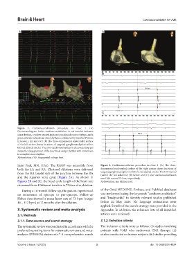
Figure 1. Cardioneuroablation procedure in Case 1. (A)
Electrocardiogram before cardioneuroablation. A red asterisk indicates
sinus rhythm, a yellow asterisk indicates junctional escape rhythm, and a
green asterisk indicates an atrial rhythm as evidenced by inverted P waves
in leads II, III, and aVF. (B) The three-dimensional endocardial surface
of the left atrium shows locations of targeted ganglionated plexi within
the red dashed circles. The post-cardioneuroablation electrocardiogram
shows the disappearance of the junctional escape rhythm with conversion
to complete sinus rhythm.
Abbreviation: aVF: Augmented voltage foot.
Saint Paul, MN, USA). The RAGP was accessible from Figure 2. Cardioneuroablation procedure in Case 2. (A) The three-
both the LA and RA. Clustered ablations were delivered dimensional endocardial surface of the right atrium shows locations of
from the RA (septal side of the junction between the RA targeted ganglionated plexi within the red dashed circles. The R-R interval
(within the red solid line) (B) before and (C) after cardioneuroablation
and the superior vena cava) (Figure 2A). As shown in was 1266 ms and 754 ms, respectively.
Figures 2B and 2C, the basal cycle length of the heart rate Abbreviation: ms: Millisecond.
decreased from 1266 ms at baseline to 754 ms after ablation.
During a 16-month follow-up, the patient experienced of the Ovid MEDLINE, Embase, and PubMed databases
no recurrence of syncope or pre-syncope. Follow-up was performed using the keywords “cardioneuroablation”
Holter data showed a mean heart rate of 73 bpm (range: and “bradycardia” to identify relevant studies published
46 – 112 bpm) at 12 months after the ablation. before 20 May 2024. No language restrictions were
applied. Details of the search strategy were provided in the
3. Systematic review and meta-analysis Appendix. In addition, the reference lists of all identified
3.1. Methods articles were reviewed.
3.1.1. Data sources and search strategy 3.1.2. Selection criteria
The systematic review was conducted in accordance with the The inclusion criteria were as follows: (i) studies involving
preferred reporting items for systematic reviews and meta- patients with VMB who underwent CNA therapy; (ii)
analyses (PRISMA) statements. A comprehensive search studies conducted on human subjects; (iii) studies providing
30
Volume 3 Issue 1 (2025) 3 doi: 10.36922/bh.4824