
Page 35 - GPD-1-2
P. 35
Gene & Protein in Disease RUNX1 gene in female-related cancers
Table 2. Summary of the functions of RUNX1 gene and the phenotypes through which the functions are expressed.
Functions Phenotypes References
Hematopoiesis Transcriptional transfer is essential for the trans-differentiation of endothelial [27,66-69]
cells into functional HSPCs.
Pre-HSCs are differentiated into HSCs.
Primitive macrophages are absent, the number of diploid megakaryocytes
decreases, and primitive erythropoiesis is unusual in the absence of RUNX1.
Nociceptive sensory neurons regulation Regulates the phenotype of several nociceptors, such as the expression of [40,67-73]
thermal receptors of the TRP class.
Hair follicle development Controls the activation of HFSCs, and increases the number of adult skin cells. [73-78]
HFSCs: Hair follicle stem cells; HSCs: Hematopoietic stem cells; HSPCs: Hematopoietic stem and progenitor cells; TRP: Transient receptor potential.
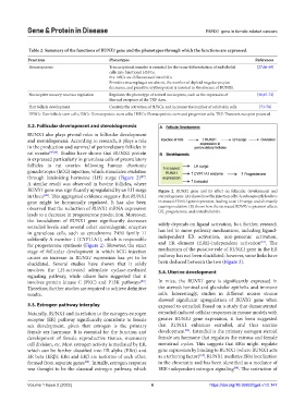
3.2. Follicular development and steroidogenesis A
RUNX1 also plays pivotal roles in follicular development
and steroidogenesis. According to research, it plays a role
in the production and survival of periovulatory follicles in
rat ovaries [85,86] . Studies have shown that RUNX1 protein B
is expressed particularly in granulosa cells of preovulatory
follicles in rat ovaries following human chorionic
gonadotropin (hCG) injection, which stimulates ovulation
through luteinizing hormone (LH) surge (Figure 2) .
[87]
A similar result was observed in bovine follicles, where
RUNX1 gene was significantly upregulated by an LH surge Figure 2. RUNX1 gene and its effect on follicular development and
in theca . This aggregated evidence suggests that RUNX1 steroidogenesis. (A) shows how the injection of hCG subsequently leads to
[88]
gene might be hormonally regulated. It has also been increased RUNX1 gene expression, leading to an LH surge, and ultimately
observed that the reduction of RUNX1 mRNA expression causing ovulation. (B) shows how the increased RUNX1 expression affects
LH, progesterone, and estradiol levels.
leads to a decrease in progesterone production. Moreover,
the knockdown of RUNX1 gene significantly decreases solely depends on ligand activation, but, further, research
estradiol levels and several other steroidogenic enzymes
in granulosa cells, such as cytochrome P450 family 11 has led to more pathway mechanisms, including ligand-
subfamily A member 1 (CYP11A1), which is responsible independent ER activation, non-genomic activation,
[91]
for progesterone synthesis (Figure 2). However, the exact and ER element (ERE)-independent activation . The
stage of follicular development in which hCG injection mechanism of the putative role of RUNX1 gene in the ER
causes an increase in RUNX1 expression has yet to be pathway has not been elucidated; however, some links have
elucidated. Several studies have shown that it solely been deduced between the two (Figure 3).
involves the LH-activated adenylate cyclase-mediated 3.4. Uterine development
signaling pathway, while others have suggested that it
involves protein kinase C (PKC) and P13K pathways . In mice, the RUNX1 gene is significantly expressed in
[89]
Therefore, further studies are required to achieve definitive the uterus’s luminal and glandular epithelia and immune
results. cells. Interestingly, studies in different mouse strains
showed significant upregulation of RUNX1 gene when
3.3. Estrogen pathway interplay exposed to estradiol. Based on a study that demonstrated
Naturally, RUNX1 and its relation to the estrogen-estrogen estradiol-induced cellular responses in mouse models with
receptor (ER) pathway significantly contribute to female greater RUNX1 gene expression, it has been suggested
sex development, given that estrogen is the primary that RUNX1 enhances estradiol, and thus uterine
[92]
female sex hormone. It is essential for the function and development . Estradiol is the primary estrogen steroid
development of female reproductive tissues, mammary female sex hormone that regulates the estrous and female
cell division, etc. Most estrogen activity is mediated by ER, menstrual cycles. This suggests that ERα might regulate
which can be further classified into ER alpha (ERα) and gene expression by binding to RUNX1 (where RUNX1 acts
[93]
ER beta (ERβ); ERɑ and ERβ are isoforms of each other, as a tethering factor) . RUNX1 mediates ERα localization
formed from separate genes . Initially, estrogen response in the chromatin and has been identified as a mediator of
[90]
was thought to be the classical estrogen pathway, which ERE-independent estrogen signaling . The restriction of
[94]
Volume 1 Issue 2 (2022) 6 https://doi.org/10.36922/gpd.v1i2.147