
Page 28 - GPD-2-2
P. 28
Gene & Protein in Disease Hematoma clearance by microglia after ICH
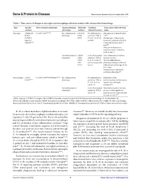
Table 1. Time course of changes in microglia and macrophage activation markers after intracerebral hemorrhage
Type Specific Flow cytometry phenotype Immunostaining Molecular Cytokines Time course with activation
markers phenotype markers released in serum status
Microglia TMEM119 CD11b CD45 int[19,20] Pro-inflammatory CD16/32 Pro-inflammatory Microglia are activated within
+
FCRLS phenotype CD86 cytokines: TNF-α, 1 h .
[42]
OLFML3 iNOS IL-6, IL-12, IL1-β, Multiple pro-inflammatory
Siglec-H and NO factors are released in the first
GPR34 [32-34] 3 days and decrease after 3
days .
[24]
Most pro-inflammatory
cytokines return to baseline
level on day 14.
Anti-inflammatory CD206 Anti-inflammatory Anti-inflammatory markers
phenotype ARG1 cytokines: TGF-β, increase on day 1.
CD36 IL-4, IL-10, IL-13, The pro-inflammatory
YM1 and various growth phenotype changes to the anti-
FIZZ1 factors inflammatory phenotype in the
first 7 days.
Predominance: alternative
activation phenotype .
[43]
Macrophages CD163 CD11b CD45 high F4/80 Ly6c [19-21] Pro-inflammatory Pro-inflammatory Infiltration of the
+
+
CD44 [34-36] phenotype cytokines: TNF-α, perihematomal and hematoma
IL-1, IL-6, IL-8, regions after microglia
and IL-12 activation .
[24]
Anti-inflammatory Anti-inflammatory Polarization markers increase
phenotype cytokines: TGF-β on day 1 with a significant
and IL-10 increase on day 3 and a further
[19]
increase on day 7 .
ARG1: Arginase 1; FCRLS: Fc receptor-like S; GPR34: G protein-coupled receptor 34; ICH: Intracerebral hemorrhage; IL-10: Interleukin 10;
iNOS: inducible nitric oxide synthase; MMΦ: Microglia/macrophages; NO: Nitric oxide; OLFML3: Olfactomedin-like 3; Siglec-H: Sialic acid binding
immunoglobulin-like lectin H; TGF-β: Transforming growth factor beta; TMEM119: Transmembrane protein 119; TNF-α: Tumor necrosis factor alpha
IL-10, a critical modulator of glial activation, is secreted clearance . However, we should identify the downstream
[45]
by monocyte-derived macrophages, resident microglia, and target molecules of CD36 in this signaling pathway.
regulatory T cells (Tregs) in the CNS. The IL-10 secreted by Exogenous administered IL-10 can inhibit apoptosis in
macrophages inhibits the activation of adjacent macrophages brain tissues around the hematoma after ICH by inhibiting
and the production of pro-inflammatory cytokines. Tregs the expression of nerve growth factor precursor (proNGF)
control immune homeostasis, suppress pro-inflammatory and p75 NTR , increasing the level of B-cell lymphoma 2
function, and promote their own immune activity through (BCL2), and decreasing the level of BCL-2-associated X
IL-10 production . Our recent research focuses on the protein (BAX), thus exerting neuroprotective effects .
[55]
[56]
IL-10 released by microglia, which maintains the balance However, we have used transmission electron microscopy
between pro- and anti-inflammatory cytokine levels [11,23] . and revealed that ferroptosis, rather than apoptosis, is the
In a mouse brain affected by ICH, IL-10 increased on day primary form of cell death after ICH [37,57] . Therefore, whether
1, peaked on day 7, and returned to baseline 14 days after endogenous and exogenous IL-10 can inhibit ferroptosis
ictus . IL-10-induced alternative microglial activation is after ICH remains unknown from a scientific standpoint.
[45]
a primary protective mechanism that modulates microglial
phagocytosis and accelerates hematoma clearance [8,26] . In our recent study, we investigated the role of IL-10 in
phagocytosis and hematoma clearance using hippocampal
Furthermore, our research has demonstrated that IL-10 slice cultures and a mouse ICH model, respectively . Our
[45]
increases the level and accumulation of phosphorylated data showed that in slice cultures, exposure to hemoglobin
STAT3 in the nucleus of Hb-treated primary microglia . increases the level of IL-10 in microglia and improves
[45]
The IL-10 signaling pathway activates STAT3, promotes phagocytosis dependent on the expression of CD36
CD36 transcription and translation, and strengthens regulated by IL-10. After ICH, IL-10 knockout mice showed
microglial phagocytosis, leading to enhanced hematoma increased iron deposition, inflammation, brain edema, and
Volume 2 Issue 2 (2023) 4 https://doi.org/10.36922/gpd.336