
Page 60 - GPD-2-2
P. 60
Gene & Protein in Disease Transfection methods for TK6 cells
A A
B
B
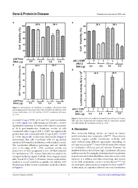
Figure 4. Optimization of conditions to minimize cell toxicity while
maximizing green fluorescent protein (GFP) transfection efficiency of the
Amaxa Nucleofector. (A) Cell viability. (B) GFP transfection efficiency.
*p < 0.05.
Figure 5. Optimization of conditions for transfection efficiency of Amaxa.
received 0.5 µg of DNA at 24 and 36 h post-transfection TK6 cells were transfected and combined with SF proprietary reagent.
(p < 0.05, Figure 5A). Cells transfected with pNL 1.1CMV (A) Cell viability. (B) Luciferase activity. *p < 0.05.
significantly expressed luciferase with maximal expression
at 36 h post-transfection. Luciferase activity of cells 4. Discussion
transfected with 0.4 µg of pNL 1.1CMV was significantly
greater than cells transfected with 0.5 µg of pNL 1.1CMV Most molecular biology studies are based on nucleic
(p < 0.05, Figure 5B). Collectively, these results (Figure 5) acid transfection into eukaryotic cells [45,46] . These studies,
demonstrate that cells transfected with 0.4 µg yielded therefore, require suitable transfection methods, with
maximal GFP transfection efficiency with minimal toxicity. each method using different approaches depending on
[47]
The transfection efficiency percentage and cell viability cell type and purpose . Every method varies with respect
were in the range of 80 – 85%. Luciferase activity was to transfection efficiency and cell toxicity. However, the
about 5.4 × 10 RLU, as opposed to 1.8 × 10 RLU obtained method of choice should have high transfection efficiency
5
5
for lipofectamine LTX. Amaxa Nucleofection yielded the and low toxicity. For example, lentivirus-based transfection
strongest luciferase signal of all the three reagents tested is an efficient method for the delivery of nucleic acids to cells;
after 24 and 36 h (Table 1). However, Amaxa nucleofection however, it is tedious, and time-consuming, and toxicity
resulted in overall weak but acceptable cell viability. GFP of the viral components can be a serious barrier [13,14] . For
fluorescence of the various transfection methods is shown the most part, optimization is a requisite for best results .
[48]
in Figure 6. In this study, we exploited different non-viral transfection
Volume 2 Issue 2 (2023) 7 https://doi.org/10.36922/gpd.0353