
Page 76 - GPD-2-4
P. 76
Gene & Protein in Disease Carpenter bee a substrate for green synthesis
at a resolution of 1 nm at room temperature. Furthermore, A B
the biosynthesized nanoparticles were characterized using
a scanning electron microscope (SEM) (JEOL JSM-IT800
HL, JEOL Ltd, Japan) at the Joint School of Nanoscience
and Nanoengineering, University of North Carolina at
Greensboro and North Carolina A and T State University,
Greensboro, North Carolina, USA.
2.3. Antimicrobial activity
The antimicrobial activity of biologically synthesized silver
nanoparticles was evaluated using the broth microdilution
method [29,33] , a simple method employed to determine
the minimum inhibitory concentration. The antibacterial
activity of the biosynthesized nanoparticles was evaluated
against common pathogenic strains of both Gram-positive
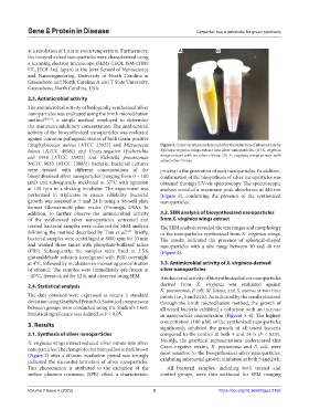
(Staphylococcus aureus [ATCC 25923] and Micrococcus Figure 2. Color variations before and after the reduction of silver nitrate by
luteus [ATCC 4698]) and Gram-negative (Escherichia Xylocopa virginica wings extract into silver nanoparticles. (A) X. virginica
coli 1946 [ATCC 25922] and Klebsiella pneumoniae wings extract with no silver nitrate. (B) X. virginica wings extract with
added silver nitrate.
NCTC 9633 [ATCC 13883]) bacteria. Bacterial cultures
were treated with different concentrations of the process in the generation of such nanoparticles. In addition,
biosynthesized silver nanoparticles (ranging from 0 – 100 confirmation of the biosynthesis of silver nanoparticles was
µM) and subsequently incubated at 37°C with agitation obtained through UV-vis spectroscopy. The spectroscopic
at 120 rpm in a shaking incubator. The experiment was analysis revealed a maximum peak absorbance at 440 nm
performed in triplicates to ensure reliability. Bacterial (Figure 3), confirming the presence of the synthesized
growth was assessed at 5 and 24 h using a 98-well plate nanoparticles.
format Glomaxmulti plate reader (Promega, USA). In
addition, to further observe the antimicrobial activity 3.2. SEM analysis of biosynthesized nanoparticles
of the synthesized silver nanoparticles, untreated and from X. virginica wings extract
treated bacterial samples were collected for SEM analysis The SEM analysis revealed the size ranges and morphology
following the method described by Tian et al. [34]. Briefly, of the nanoparticles synthesized from X. virginica wings.
bacterial samples were centrifuged at 8000 rpm for 10 min The results indicated the presence of spherical-shaped
and washed three times with phosphate-buffered saline nanoparticles with a size range between 10 and 40 nm
(PBS). Subsequently, the samples were fixed in 2.5% (Figure 4).
glutaraldehyde solution (configured with PBS) overnight
at 4°C, followed by incubation in increasing concentration 3.3. Antimicrobial activity of X. virginica-derived
of ethanol. The samples were immediately pre-frozen at silver nanoparticles
−20°C, freeze-dried for 12 h, and observed using SEM. Antibacterial activity of biosynthesized silver nanoparticles
2.4. Statistical analysis derived from X. virginica was evaluated against
K. pneumonia, E coli, M. luteus, and S. aureus at two time
The data obtained were expressed as means ± standard points (i.e., 5 and 24 h). As indicated by the results obtained
deviation using GraphPad Prism 8.0. Statistical comparisons through the broth microdilution method, the growth of
between groups were conducted using the Student’s t-test. all tested bacteria exhibited a reduction with an increase
Statistical significance was defined as P < 0.05. in nanoparticle concentration (Figures 5–8). The highest
3. Results concentration (100 µM) of the synthesized nanoparticles
significantly inhibited the growth of all tested bacteria
3.1. Synthesis of silver nanoparticles compared to the control at both 5 and 24 h (P < 0.05).
X. virginica wings extract reduced silver nitrate into silver Notably, the graphical representations underscored that
nanoparticles. The change of color from yellow to dark brown Gram-negative strains, K. pneumonia and E. coli, were
(Figure 2) after a 60-min incubation period was strongly most sensitive to the biosynthesized silver nanoparticles,
indicated the successful formation of silver nanoparticles. exhibiting substantial growth inhibition at both 5 and 24 h.
This phenomenon is attributed to the excitation of the All bacterial samples, including both treated and
surface plasmon resonance (SPR) effect, a characteristic control groups, were thin sectioned for SEM imaging
Volume 2 Issue 4 (2023) 3 https://doi.org/10.36922/gpd.2155