
Page 79 - GPD-2-4
P. 79
Gene & Protein in Disease Carpenter bee a substrate for green synthesis
A B
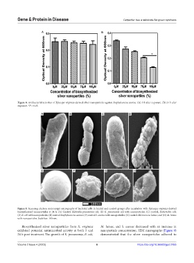
Figure 8. Antibacterial activities of Xylocopa virginica-derived silver nanoparticles against Staphylococcus aureus. (A) 5 h after exposure. (B) 24 h after
exposure. *P < 0.05.
A B C D
E F G H
Figure 9. Scanning electron microscopy micrographs of bacterial cells in treated and control groups after incubation with Xylocopa virginica-derived
biosynthesized nanoparticles at 24 h. (A) Control Klebsiella pneumonia cell; (B) K. pneumonia cell with nanoparticles; (C) control, Escherichia coli;
(D) E. coli with nanoparticles; (E) control Staphylococcus aureus; (F) control S. aureus with nanoparticles; (G) control Micrococcus luteus; and (H) M. luteus
with nanoparticles. Scale bar: 100 nm.
Biosynthesized silver nanoparticles from X. virginica M. luteus, and S. aureus decreased with an increase in
exhibited potential antimicrobial activity at both 5 and nanoparticle concentration. SEM micrographs (Figure 9)
24 h post-treatment. The growth of K. pneumonia, E. coli, demonstrated that the silver nanoparticles adhered to
Volume 2 Issue 4 (2023) 6 https://doi.org/10.36922/gpd.2155