
Page 140 - GPD-4-1
P. 140
Gene & Protein in Disease Rotavirus diversity in Uttar Pradesh
Table 1. Primer pairs used for detection and characterization of group A rotaviruses
Rotavirus A Primer Sequence (5’ to 3’) Size (bp) Annealing References
gene segments Temperature (°C)
VP4 (P) Con3F TGGCTTCGCTCATTTATAGACA 877 53 41
Con2R ATTTCGGACCATTTATAACC
Wang F20 TGGCTTCGCTCATTTATATAFAC 1185 53 42
Wang 1185R GACTGGCCATGCACCTACAGGT
VP7 (G) 9con1F TAGCTCCTTTTAATGTATGG 1030 52 43
9con2R GTATAAAATACTTGCCACCA
Vp7-F59 GCTCCTTTTAATGTATGGTAT 960 54.5 44
Vp7-R998 ARTGAYCKTGATCKTTTGGACAT
TNG C1-F GGCTTTAAAAGAGAGAATTTCCGTC TGG 1065 52.5 45
TNG C2-R GGTCACATCATACAATTCTAATCTAAG
VP6 (I) Gen VP6 F GGCTTTWAAACGAAGTCTTC 1340 48 3
Gen VP6 R GGTCACATCCTCTCACT
Ghosh VP6 F GGCTTTAAAACGAAGTCTTC 1340 52 46
Ghosh VP6 R GGTCACATCCTCTCACT
NSP4 (E) Gen Nsp4 F Nsp4 722R TAAAAGTTCTGTTCCGAGAGAG 722 52 13
TTAAGACCGTTCCTTCCATT
3. Results
3.1. Incidence of RVA
RVA is identified by its characteristic 11-banded pattern
(4:2:3:2) on RNA-PAGE, whereas amplification of the VP6
gene is confirmed by the production of 1340 bp amplicons
using RT-PCR. Out of the 100 stool samples from children,
32 tested positive for RVA, whereas the remaining 68 did
not yield any RV identification profiles through either
method. Four RNA-PAGE-positive samples failed to be
amplified using RT-PCR. Among the 32 RVA-positive
samples, 21 (65.63%) could be detected by RNA-PAGE,
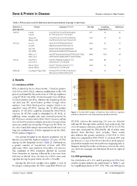
whereas 28 (87.5%) could be identified by RT-PCR. In Figure 1. Positive field samples of Rotavirus show different migration
patterns in ribonucleic acid-polyacrylamide gel electrophoresis.
addition, seven samples that were detected positive by
RT-PCR were not detected by RNA-PAGE. However, all but
four of the RVA-positive samples identified through PAGE RT-PCR, whereas the remaining 120 were not detected
were also confirmed to have the VP6 gene by RT-PCR. On with any RV through either method used in the study. Out
the other hand, field samples demonstrated both short and of the 80 samples detected positive by RT-PCR, 51 (37.5%)
long electropherotypes of RNA segregation in the RNA- were also confirmed by RNA-PAGE, all of which were
PAGE analysis (Figure 1). derived from diarrheal stool samples. These results
also indicate the greater sensitivity of RT-PCR in RVA
One private hospital in the densely populated city of detection compared to RNA-PAGE. In contrast, RVA was
Bareilly recorded the highest incidence of cases (47.5%) not detected in any of the 64 non-diarrheal stool samples.
among the five hospitals studied (Table 2). Furthermore, All positive samples were sourced from a single pig farm in
a greater number of hospitalized children with RVA Rupapur during December and January, whereas the other
infections (59%) were noted in December and January. two farms in Izatnagar and Ismilepur showed no signs of
The incidence of RVA infections showed an increase RVA.
from November to January, followed by a decrease from
February to March. Thus, the majority of RVA cases were 3.2. RVA genotyping
reported during the peak winter months in Bareilly. The distribution of G, P, I, and E genotypes in RVA from
Among the 200 stool samples from piglets, a total of positive human samples are summarized in Table 3, and
80 samples tested positive for RVA using RNA-PAGE and the amplified products of VP7, VP4, VP6, and NSP4 genes
Volume 4 Issue 1 (2025) 4 doi: 10.36922/gpd.6237