
Page 141 - GPD-4-1
P. 141
Gene & Protein in Disease Rotavirus diversity in Uttar Pradesh
Table 2. Incidence of RVA in diarrheal stool samples of humans
Months (A) (B) (C) (D) (E) Total
(HBI) (HBM) (HBS) (HBG) (HBD)
PAGE RT‑PCR PAGE RT‑PCR PAGE RT‑PCR PAGE RT‑PCR PAGE RT‑PCR PAGE RT‑PCR
(%) (%) (%) (%) (%) (%) (%) (%) (%) (%) (%) (%)
November-17 1/4 2/4 0/6 0/6 0/8 0/8 0/1 0/1 0/2 0/2 1/21 2/21
December-17 3/6 4/6 1/5 2/5 1/4 1/4 1/6 0/6 2/7 3/7 8/28 10/28
January-18 5/10 6/10 1/1 1/1 0/1 0/1 0/2 0/2 1/3 2/3 7/17 9/17
February-18 3/12 4/12 0/3 0/3 0/2 0/2 0/3 1/3 1/5 2/5 4/25 6/25
March-18 1/8 1/8 0 0 0 0 0 0 0/1 0/1 1/9 1/9
Total 13/40 17/40 2/15 3/15 1/15 1/15 1/12 1/12 4/18 6/18 21/100 28/100
(32.5) (42.5) (32.5) (13.3) (6.66) (6.66) (8.33) (8.33) (22.2) (33.3) (21.0) (28.0)
RVA+Samples 19/40 3/15 1/15 2/12 7/18 32/100
(47.5) (20.0) (6.66) (16.6) (38.8) (32.0)
Notes: HBI: Hospital 1; HBM: Hospital 2; HBS: Hospital 3; HBG: Hospital 4; HBD: Hospital 5.
Abbreviation: RVA: Rotavirus A; RT-PCR: Reverse transcription polymerase chain reaction; PAGE: Polyacrylamide gel electrophoresis.
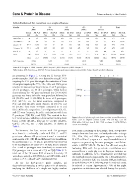
are presented in Figure 2. Among the 32 human RVA-
positive samples, 28 (87.5%) were detectable using RT-PCR
targeting the VP6 gene. Genotypic determination of these
28 samples targeting the VP7, VP4, VP6, and NSP4 genes
revealed 24 instances of G genotypes, 25 of P genotypes,
28 of I genotypes, and 27 of E genotypes. When further
characterizing the VP7 gene sequences, the G3 (46.42%)
genotype was found to be the most prevalent, followed by
G1 (28.57%) and G2 (10.71%). In terms of P genotypes,
P[8] (60.71%) was the most dominant, compared to
P[4] and P[6] (14.28% each). Besides, I1 (78.57%) and
E1 (75.0%) were more prevalent compared to I2 and
E2 (21.42% each), respectively. Three G genotypes (G1, G2,
and G3) were frequently found in combination with three
P genotypes (P[4], P[6], and P[8]). This resulted in four
G-P combinations, with the predominant circulating strain Figure 2. Result of gel electrophoresis shows amplified genes of Rotavirus A
being G3P[8] (46.42%), followed by G1P[8] (14.28%), Notes: Lane N: Negative control; Lane1: VP4 (850 bp); Lane M:
DNA ladder (100 bp); Lane 2: VP6 (1060 bp); Lane 3: NSP4 (700 bp);
G2P[4] (10.71%), and G1P[6] (14.28%), as shown in Lane 4: VP7 (900 bp).
Table 3.
Furthermore, the RVA strains with G3 genotype RVA strain circulating on the Rupapur farm. Five positive
were found to consistently coexist with P[8], I1, and E1 samples from this farm were randomly selected to undergo
genotypes, whereas G2 genotypes showed a consistent VP7, VP4, VP6, and NSP4 gene sequence analysis by
combination with P[4], I2, and E2 genotypes. In addition, BLAST and RotaC program, and they all revealed identical
the coexistence of G1, I1, and E1 genotypes was observed sequences representing the same genotypic constellation,
to be accompanied by either P[8] or P[6]. It also appears which is G9P[13]-I5-E1. The fact that all stool samples
that I2 and E2 genotypes were found only in strains with harboring RVA with this genotypic constellation were
P[4] genotype, not in those with P[6] or P[8] (Table 3). In sourced from a single pig farm in Rupapur indicates an
total, out of the 28 human RVA strains in this study, 13 outbreak. In addition, further investigation unveiled that
were characterized as G3P[8]I1E1, 4 as G1P[6]I1E1, and 3 the diarrheal episodes began at the end of November 2017,
each as G1P[8]I1E1 and G2P[4]I2E2 genotypes. peaked in December 2017 and January 2018, and subsided
As for the RVA-positive piglet samples, gel by early February 2018, inferring that the outbreak could
electrophoresis revealed a similar pattern for all samples, be related to winter. Approximately 75% of the piglet
implying the likelihood of a single and highly virulent population on this farm was affected during this episode,
Volume 4 Issue 1 (2025) 5 doi: 10.36922/gpd.6237