
Page 132 - GPD-4-2
P. 132
Gene & Protein in Disease Binding of 11q to DENV and WNV proteases
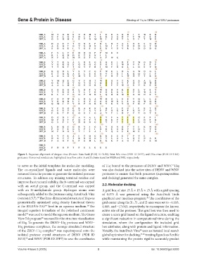
Figure 1. Sequence alignment of dengue virus (Protein Data Bank [PDB] ID 3U1I), West Nile virus (PDB ID 2FP7), and Zika virus (PDB ID 5H4I)
proteases. Conserved residues are highlighted in yellow color. A and B chains stand for NS2B and NS3, respectively.
13
to serve as the initial templates for molecular modeling. of 11q bound to the proteases of DENV and WNV. 11q
The co-crystallized ligands and water molecules were was also docked into the active sites of DENV and WNV
removed from the protein to generate the isolated protease proteases to ensure that both processes (superimposition
structures. To address any missing terminal residue and and docking) generated the same complex.
improve the structural stability, the N-terminal was capped
with an acetyl group, and the C-terminal was capped 2.2. Molecular docking
with an N-methylamide group. Hydrogen atoms were A grid box of size 15 Å × 15 Å × 15 Å with a grid spacing
subsequently added to the protease using AutoDock Vina of 0.375 Å was generated using the AutoDock Tools
38
(version 1.5.7). The three-dimensional structure of 11q was graphical user interface program. The coordinates of the
38
geometrically optimized using density functional theory grid center along the X-, Y-, and Z-axes were set to −4.183,
at the B3LYP/6-31G** level in an aqueous medium. The 4.685, and −17.043, respectively, to encompass the known
34
integral equation formalism of the polarized continuum active site of the protease. This grid box was then used to
model was used to model the aqueous medium. The Gauss create a score grid based on the ligand structure, enabling
39
View 5.0 program was used for the structure visualisation a significant reduction in computational time during the
40
of 11q. To generate the DENV–11q protease and WNV– simulation, where the configuration file included grid
11q protease complexes, the average simulated structure box attributes, along with protein and ligand information.
of the ZIKV–11q complex was superimposed onto the Notably, the AutoDock Vina uses an iterated local search
34
38
isolated protease crystal structures of DENV (PDB ID global optimizer for docking, 41,42 treating ligands as flexible
3U1I) and WNV (PDB ID 2FP7) to save the coordinates while maintaining the protein rigid to accurately predict
12
Volume 4 Issue 2 (2025) 3 doi: 10.36922/gpd.8293