
Page 24 - GTM-2-2
P. 24
Global Translational Medicine Use of cardio biomarker in diagnosis of AMI
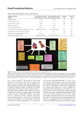
Table 1. Biomarker for diagnosis of myocardial infarction
Diagnostic indicator Time required for marker Time required for marker Specificity Sensitivity
(marker) level to peak after onset (h) level to peak after onset (h) (%) (%)
Myoglobin (MYO) 4–6 20–25 80 92
Cardiac troponin I (cTnI) 12–24 5–9 98.4 98.7
Cardiac troponin T (cTnT) 18–36 5–14 100 99
Creatinine kinase (CK) 20–30 24–48 91 24
Glucose dehydrogenase (GDH) 48–72 10–15 - -
N-terminal pro-B-type natriuretic peptide (NT-pro-BNP) No clinical consensus No clinical consensus 72.6 50.7
Heart-type fatty acid-binding protein (H-FABP) 6 0.5–1 68 89.7
Creatine kinase-myoglobin binding (CK-MB) 12–24 3–4 92 44.8
B-type natriuretic peptide (BNP) No clinical consensus No clinical consensus 90 86
Figure 2. Therapeutical potential and functional role of biomarkers of acute myocardial infarction.
Abbreviations: AST: Aspartate transaminase; BNP: B-type natriuretic peptide; CK-MB: Creatine kinase-myoglobin binding; Copeptin: C-terminal pro-arginine
vasopressin; CRP: C-reactive protein; Cys-C: Cystatin C; Gal-3: Galectin-3; GDF-15: Growth differentiation factor-15; H-FABP: Heart-type fatty acid-binding
protein; HF: Heart failure; HFpEF: Heart failure with preserved ejection fraction; IL: Interleukin; MR-pro-ANP: Midregional pro-atrial natriuretic peptide; NP:
Natriuretic peptide; NT-pro-BNP: N-terminal pro-B-type natriuretic peptide; PLT: Platelet; SP-1: Specificity protein 1; ST-2: suppression of tumorigenicity 2.
into a colorful product . The process for ELISA-based color measured spectrophotometrically . In 2010, Kim et
[58]
[57]
colorimetric immunoassays often entails covering a solid al. developed a test of lateral flow strips comprising a sample
surface (such as a microplate) with a capture antibody or an pad, conjugate pad, nitrocellulose membrane, and absorbent
antigen specific to the target analyte. After blocking to reduce pad. After the cTnl antigen is added to the sampling pad,
non-specific binding, the sample is introduced to let the target two separate gold nanoparticles (AuNP) conjugates are
analyte attach to the antigen or antibody that will capture it. deposited onto the conjugated pad and then flow out. First,
The analyte’s distinct epitope is then bound by an enzyme- cTnI reacts with AuNP containing anti-troponin antibodies
labeled detecting antibody or antigen, which is then added. I, followed by a second AuNP interacting with the first AuNP
A substrate solution is added after incubation and washing by BSA and anti-BSA antibody interactions. This method
steps are performed to remove unbound components. shows higher signal intensity than the conventional lateral
An enzyme label catalyzes a reaction with a substrate that flow assay (LFA) method, and after passing through the NC
results in the production of a colored product. The analyte membrane, conjugated AuNPs were immobilized onto the
concentration is directly proportional to the intensity of surface and finally, by dipping the cut LFA strip into troponin
Volume 2 Issue 2 (2023) 6 https://doi.org/10.36922/gtm.0403