
Page 61 - GTM-3-1
P. 61
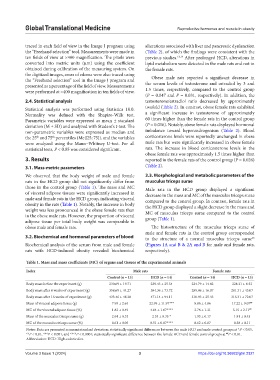
Global Translational Medicine                                   Reproductive hormones and muscle in obesity


            traced in each field of view in the Image J program using   alterations associated with liver and pancreatic dysfunction
            the “Freehand selection” tool. Measurements were made in   (Table 2), of which the findings were consistent with the
            ten fields of view at ×400 magnification. The pixels were   previous studies. 13,14  After prolonged HCD, alterations in
            converted  into  metric  units  (μm)  using  the  coefficient   lipid metabolism were detected in the male rats and not in
            obtained during calibration of the measuring system. On   the female rats.
            the digitized images, areas of edema were also traced using   Obese male rats reported a significant decrease in
            the “Freehand selection” tool in the Image J program and
            presented as a percentage of the field of view. Measurements   the serum levels of testosterone and estradiol by 3 and
            were performed at ×400 magnification in ten fields of view.  1.5  times, respectively, compared to the control group
                                                               (P = 0.047 and P = 0.031, respectively). In addition, the
            2.4. Statistical analysis                          testosterone/estradiol  ratio decreased by  approximately
            Statistical analysis was performed using Statistica 10.0.   twofold (Table 2). In contrast, obese female rats exhibited
            Normality was  defined  with  the  Shapiro-Wilk  test.   a significant increase in testosterone of approximately
            Parametric variables were expressed as mean ± standard   60 times higher than the female rats in the control group
            deviation (М ± SD) and analyzed with Student’s t-test. The   (P = 0.016). Notably, obese female rats displayed hormonal
            non-parametric  variables  were  expressed  as median and   imbalance toward hyperandrogenism (Table  2). Blood
            the 25  and 75  percentiles (Me [25; 75]), and the variables   corticosterone levels were reportedly unchanged in obese
                       th
                 th
            were analyzed using the Mann–Whitney U-test. For all   male rats but were significantly increased in obese female
            statistical tests, P < 0.05 was considered significant.  rats.  The  increase  in  blood  corticosterone  levels in  the
                                                               obese female rats was approximately 1.5 times higher than
            3. Results                                         reported in the female rats of the control group (P = 0.026)
            3.1. Mass-metric parameters                        (Table 2).
            We observed that the body weight of male and female   3.3. Morphological and metabolic parameters of the
            rats in the HCD group did not significantly differ from   musculus triceps surae
            those in the control group (Table 1). The mass and MC   Male rats in the HCD group displayed a significant
            of visceral adipose tissues were significantly increased in   decrease in the mass and MC of the musculus triceps surae
            male and female rats in the HCD group, indicating visceral   compared to the control group. In contrast, female rats in
            obesity in the rats (Table 1). Notably, the increase in body   the HCD group displayed a slight decrease in the mass and
            weight was less pronounced in the obese female rats than   MC  of musculus triceps  surae  compared  to  the control
            in the obese male rats. However, the proportion of visceral   group (Table 1).
            adipose tissue per total body weight was comparable in
            obese male and female rats.                          The histostructure of the musculus triceps surae of
                                                               male and female rats in the control group corresponded
            3.2. Biochemical and hormonal parameters of blood  to  the  structure  of  a  normal  musculus  triceps  surae
                                                                                                            15
            Biochemical analysis of the serum from male and female   (Figures 1А and В & 2А and В for male and female rats,
            rats with HCD-induced obesity revealed biochemical   respectively).

            Table 1. Mass and mass coefficients (MC) of organs and tissues of the experimental animals
            Index                                         Male rats                        Female rats
                                               Control (n = 13)  HCD (n = 14)     Control (n = 14)  HCD (n = 13)
            Body mass before the experiment (g)  230.69 ± 19.71  229.43 ± 25.54    224.79 ± 14.82   228.31 ± 6.52
            Body mass after 8 weeks of experiment (g)  358.69 ± 44.27  384.36 ± 73.72  289.86 ± 16.07  281.31 ± 43.07
            Body mass after 16 weeks of experiment (g)  433.46 ± 48.38  471.14 ± 94.11  310.93 ± 25.43  316.31 ± 72.07
            Mass of visceral adipose tissue (g)  7.99 ± 2.64     22.99 ± 11.97***   8.86 ± 4.86     17.22 ± 9.07 ##
            MC of the visceral adipose tissue (%)  1.82 ± 0.49   4.64 ± 1.67****    2.78 ± 1.31     5.15 ± 2.11 ##
            Mass of the musculus triceps surae (g)  2.64 ± 0.33   2.34 ± 0.35 *     1.93 ± 0.17      1.81 ± 0.45
            MC of the musculus triceps surae (%)  0.61 ± 0.05    0.51 ± 0.07****    0.62 ± 0.07      0.58 ± 0.11
            Notes: Data are presented as mean±standard deviation; statistically significant differences between the male HCD and male control groups at *P < 0.05,
            **P < 0.01, ***P < 0.001, and ****P < 0.0001; statistically significant difference between the female HCD and female control groups at  P < 0.01.
                                                                                                 ##
            Abbreviation: HCD: High-calorie diet.

            Volume 3 Issue 1 (2024)                         3                        https://doi.org/10.36922/gtm.2321